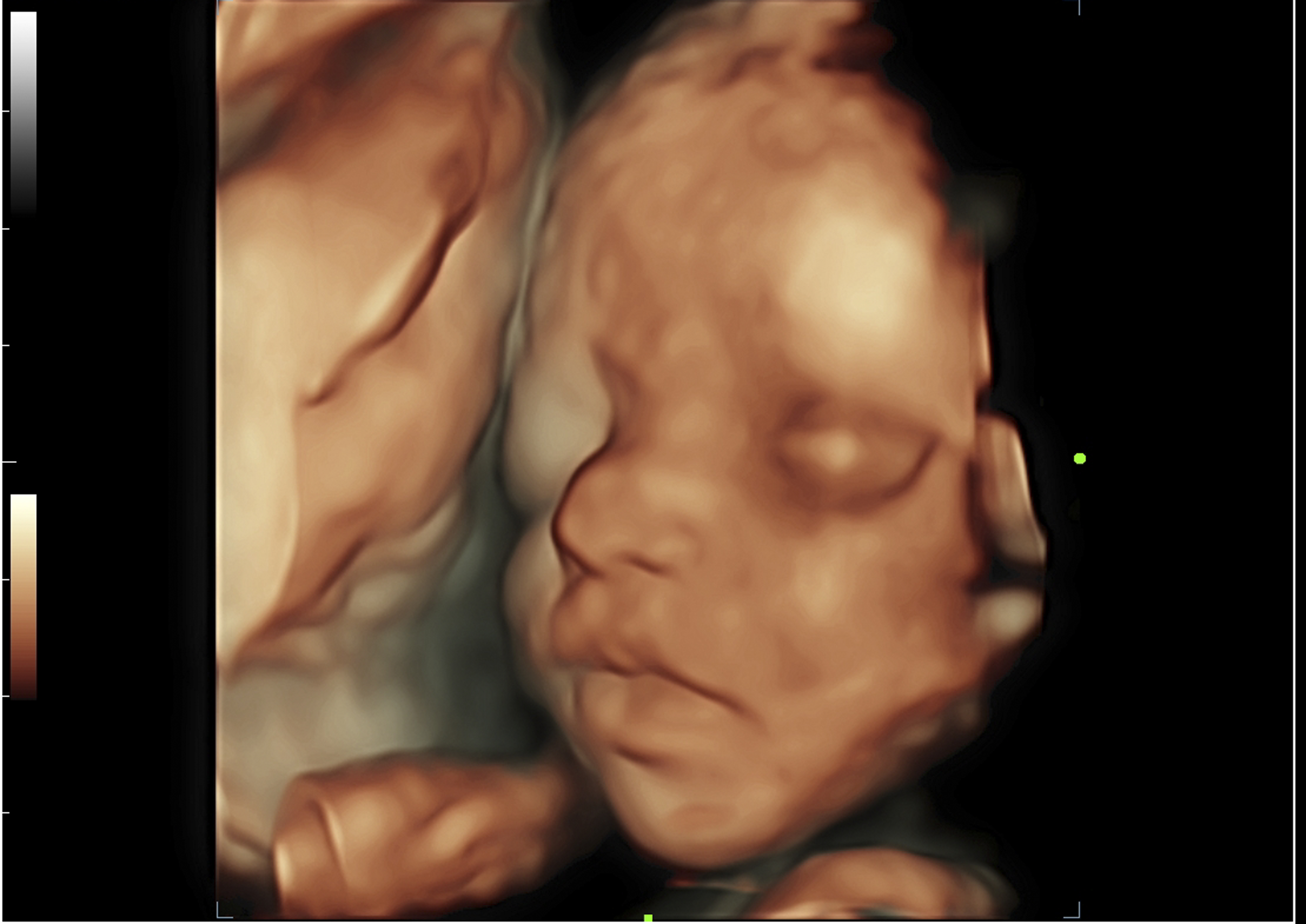
3D/4D Obestrics Scan
Ultrasound scans including 3D and 4D are more like a real-time photograph of your baby. With 3D scanning, many pictures of the child are taken in 2D and then merged to create a 3D image effect. With 4D scanning, pictures are made in real time and you can see what your child is doing in your womb at the time, such as moving of legs and arms or opening of the eyes.
Ultrasound scans are an essential tool to examine the health and internal organs of a growing fetus. It helps the gynaecologists to identify any complications. This identification allows doctors at Motherhood Hospitals to treat the baby at the earliest. These 3D/4D scans done throughout pregnancy, help the experts to detect any kind of anomalies such as a cleft lip, underdeveloped limbs helps you keep a track of spinal problems and other congenital disabilities. It also helps in monitoring the amniotic fluid.
Benefits of 3D Scan
- It has a better view of foetal heart structures because it produces images that cannot be achieved through 2D models
- It helps to diagnose neural defects and foetal musculoskeletal
- It helps to diagnose foetal face defects such as a cleft lip
- It takes less time to get the standard view of the plane
- The recorded volume data can be kept on record for better diagnosis and expert advice
- It is easy to study and understand
Benefits of 4D Scan
- Increase parenting relationships with the child
- Shorter time for foetal heart screening and diagnosis
- The recorded data can be kept on record for expert review
- It also shares the advantages of 3D ultrasound including examining the foetal heartbeat, placental localization and the assessment of foetal well being and growth
How does 3D/4D scan work?
The best sonographer will help you with the complete procedure. A probe or a transducer coated with a conductive gel is a device used to perform the ultrasound which sends a signal inside the womb. 3D ultrasounds create a three-dimensional image, while 4D ultrasounds create a live video effect, you can watch the hands moving, legs moving, smiling, yawning of the foetal. Both the scans are safe and painless.

