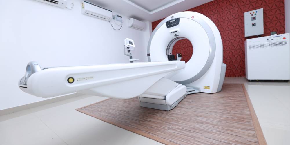
COVID-19 DIAGNOSIS USING CT SCAN
Omicron is a variant of SARS-CoV-2 that has been identified initially in COVID-19 patients in Botswana and South Africa. Studies suggest that symptoms produced by omicron are less severe than those caused by previously identified variants, and those people recorded as infected with the Omicron variant are about 15-20% less likely to seek hospital care and 40% less likely to be admitted for a night or longer than those with Delta. Along with other laboratory testings, chest CT scans also serves to be helpful to diagnose Omicron COVID-19 in individuals with a high clinical suspicion of infection.
Advantages of multi-slice CT scan
- The image quality is way much better than its traditional counterpart.
- Better image quality results in the early and precise diagnosis of diseases, which eventually means saving a lot of suffering and money.
- The scanners used work at very high speed. Where it takes around 10 minutes on a conventional single-slice CT to do a scan, the multi scanner can do the job within a few seconds and even more efficiently.
- Due to the less time taken, the multi-slice CT scan can be done on people in restless stage or those who are unconscious. This is also helpful with children as it is difficult to get them to lie in one position for a long time.
- The emission of radiation is very low. A single slice scanner emits more harmful rays as compared to the multi-slice scanner. For this reason, a multi-slice scan is favoured for children.
- In some cases of a single slice, the disease remains undiagnosed in its early stages due to low image quality. Later on, when the difficulties are aggravated, the patient is referred for high-quality scans and sometimes so much time is lost that the diseases go untreatable.
- Non-invasive Angiography can only be done on an MSCT. In angiography, multi-slice CT helps in creating a 3D image with precise visualization of arteries & veins.
- 3D images of the bones, rib cage, spine etc can be obtained in multislice CT scan which results in better assessment of the fractures and gives better images for the surgeon to operate upon the patient.
- It has been found in multiple studies that multislice CT leads to less repeatability as the images are good to diagnose in only one scan. This avoids unnecessary cost and radiation to the patient.

