
3D/4D Obstetric Scan in OMR
Pregnancy Ultrasound at AVR Scan Centre
Pregnancy is a time when you naturally want to understand how your baby is developing and what each scan means. If your doctor has recommended an obstetric ultrasound or 3D/4D pregnancy scan in OMR, you may have questions about what the scan examines, when it may be performed, how to prepare and whether ultrasound is safe during pregnancy.
Obstetric ultrasound uses sound waves to create images of the developing fetus and pregnancy-related structures. It can help healthcare professionals assess aspects of pregnancy such as fetal development, growth, position, placenta, amniotic fluid and other findings depending on the examination. Ultrasound does not use ionizing radiation.
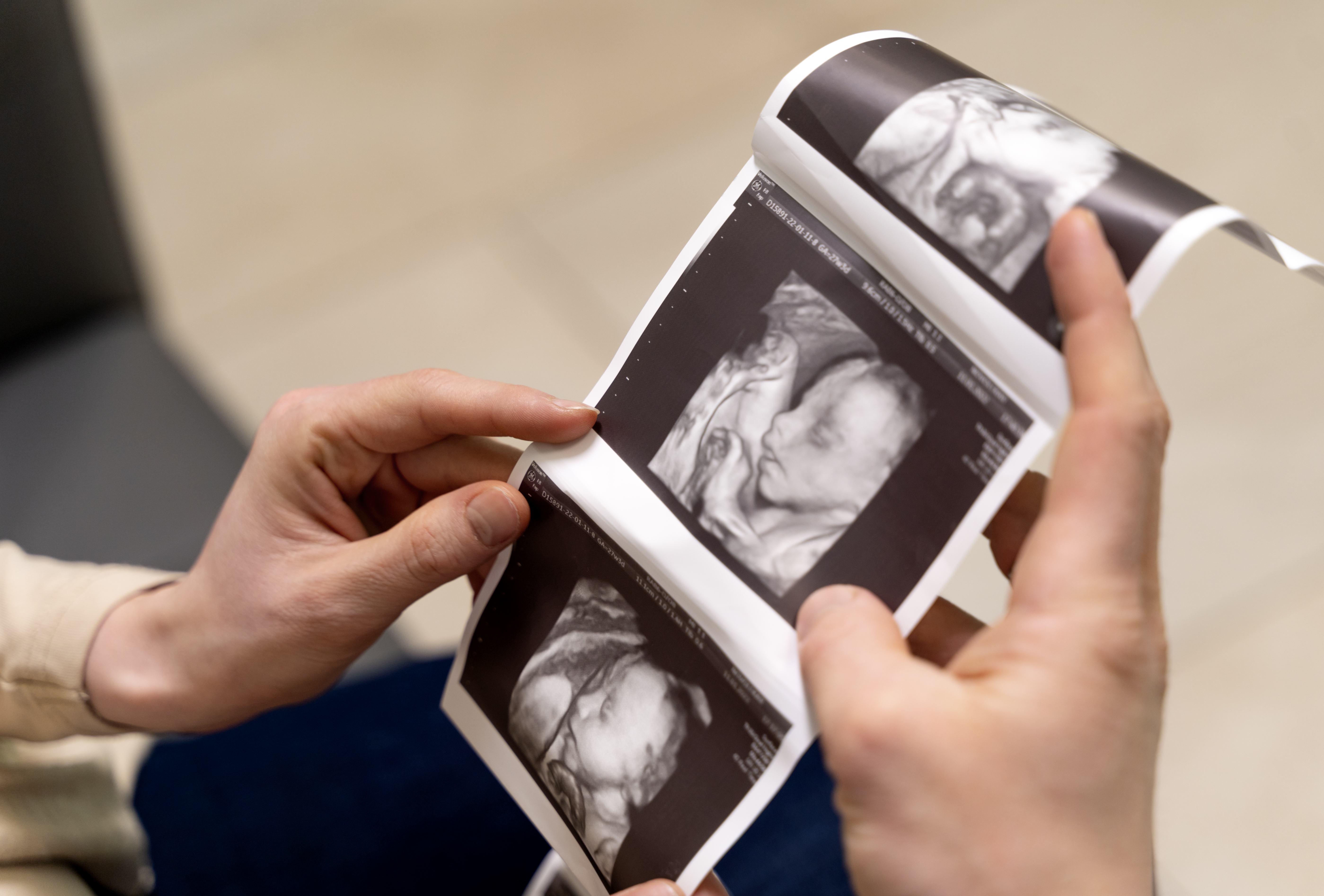
3D ultrasound creates three-dimensional images from ultrasound data, while 4D ultrasound adds real-time movement to 3D imaging. The usefulness of 3D or 4D imaging depends on the clinical indication, gestational age, fetal position and other imaging factors.
AVR Scan Centre provides 3D/4D Obstetric Scan services for patients looking for pregnancy imaging in and around the OMR region.
What Is a 3D/4D Obstetric Scan?
A 3D/4D obstetric scan is a pregnancy ultrasound examination that can create three-dimensional images of the fetus, with 4D ultrasound adding the element of real-time movement.
A standard obstetric ultrasound generally produces two-dimensional images. Three-dimensional ultrasound processes ultrasound data to create a three-dimensional representation, while four-dimensional ultrasound allows those 3D images to be viewed with movement over time.
Depending on the examination and clinical purpose, obstetric ultrasound may provide information about:
- Fetal development
- Fetal growth
- Fetal position
- Fetal movement
- Fetal heart activity
- Placental location
- Amniotic fluid
- Number of fetuses
- Selected fetal anatomical structures
The exact information obtained depends on the stage of pregnancy, fetal position, examination type and clinical indication.
3D/4D Obstetric Scan in OMR – Where Can You Get a Pregnancy Scan?
If you are searching for a 3D/4D Obstetric Scan in OMR, AVR Scan Centre provides 3D/4D Obstetric Scan services for pregnancy-related diagnostic imaging.
The Sholinganallur centre serves patients looking for diagnostic imaging in the OMR region.
Patients travelling from Padur, Navalur, Siruseri, Kelambakkam, Thiruporur and nearby OMR areas should contact AVR Scan Centre before visiting to confirm the availability of the required obstetric examination.
Before your appointment, confirm:
- The type of pregnancy scan recommended
- The appropriate gestational stage for your examination
- Whether any preparation is required
- Whether a full bladder is necessary for your specific scan
- Appointment availability
- Any previous pregnancy reports that should be brought
AVR Scan Centre – Sholinganallur:
First Floor, No. 278/1 & 278/2, Commercial Building,
Ramanyam Pushkar 2, Kalaignar Karunanidhi Salai,
Sholinganallur – 600119.
Why Is an Obstetric Ultrasound Recommended During Pregnancy?
An obstetric ultrasound may be recommended to monitor pregnancy and assess the fetus and pregnancy-related structures at different stages of pregnancy.
Depending on the examination, ultrasound may help healthcare professionals assess:
- Presence and location of the pregnancy
- Gestational age
- Number of fetuses
- Fetal growth
- Fetal position
- Fetal movement
- Fetal heart activity
- Placental location
- Amniotic fluid
- Selected fetal anatomy
- Certain pregnancy-related concerns
The timing and type of ultrasound depend on the individual pregnancy and the purpose of the examination.
ACOG notes that prenatal ultrasound may be standard, limited or specialized depending on the clinical question.
What Can a 3D/4D Obstetric Scan Show?
A 3D/4D obstetric scan can provide additional views of fetal structures and movement, but the information obtained varies according to the pregnancy and examination conditions.
Fetal Anatomy
Three-dimensional imaging can provide additional perspectives of selected fetal structures when suitable images can be obtained.
Fetal Movement
4D imaging can display movement in real time, allowing dynamic visualization during the scan.
Fetal Position
The position of the fetus can affect the images obtained and may also be part of the assessment during obstetric ultrasound.
Placenta and Amniotic Fluid
Obstetric ultrasound can help assess placental position and the amount of amniotic fluid around the fetus.
Fetal Growth
Ultrasound can be used to assess fetal growth and measurements appropriate to the examination and stage of pregnancy.
What Is the Difference Between 2D, 3D and 4D Ultrasound?
The main difference is how the ultrasound information is displayed.
| Ultrasound Type | What It Provides |
|---|---|
| 2D Ultrasound | Conventional two-dimensional images |
| 3D Ultrasound | Three-dimensional images reconstructed from ultrasound data |
| 4D Ultrasound | Three-dimensional images displayed with real-time movement |
3D and 4D imaging do not replace the need for appropriate medical assessment. The examination should be selected according to the clinical purpose.
When Is a 3D/4D Pregnancy Scan Done?
There is no single gestational week that is appropriate for every 3D/4D scan.
The most suitable timing depends on:
- Purpose of the examination
- Gestational age
- Fetal position
- Amount of amniotic fluid
- Placental position
- Maternal body characteristics
- Imaging conditions
- Doctor's recommendation
A standard obstetric ultrasound may be scheduled according to routine prenatal care, while specialized examinations are performed when a specific clinical question needs to be addressed.
ACOG notes that a standard prenatal ultrasound is commonly performed around 18–22 weeks, although timing varies by clinical circumstances.
For a 3D/4D examination specifically, the visibility of the fetus can vary significantly from one pregnancy to another, so the best time should be determined according to the reason for the scan and the advice of the treating healthcare professional.
Is 3D/4D Ultrasound Safe During Pregnancy?
Diagnostic obstetric ultrasound is considered safe when appropriately performed for medical purposes by trained professionals.
Ultrasound does not use ionizing radiation. However, ultrasound energy can produce biological effects under certain exposure conditions, which is why professional organizations recommend prudent use and keeping exposure as low as reasonably achievable while obtaining the necessary diagnostic information.
The AIUM recommends that fetal ultrasound examinations be performed by appropriately trained and credentialed professionals and discourages nonmedical or unnecessary ultrasound exposure.
For this reason, 3D/4D ultrasound should be treated as a medical imaging examination rather than simply a keepsake or entertainment scan.
Does a 3D/4D Obstetric Scan Use Radiation?
No. Ultrasound does not use ionizing radiation.
Obstetric ultrasound uses high-frequency sound waves to create images of the fetus and pregnancy-related structures.
This is different from:
- X-Ray: X-rays
- CT Scan: X-rays
- MRI: Magnetic fields and radio waves
Even though ultrasound does not use ionizing radiation, it should still be used appropriately and for a medical purpose.
Is a 3D/4D Pregnancy Scan Painful?
A routine abdominal obstetric ultrasound is generally non-invasive and is usually not painful.
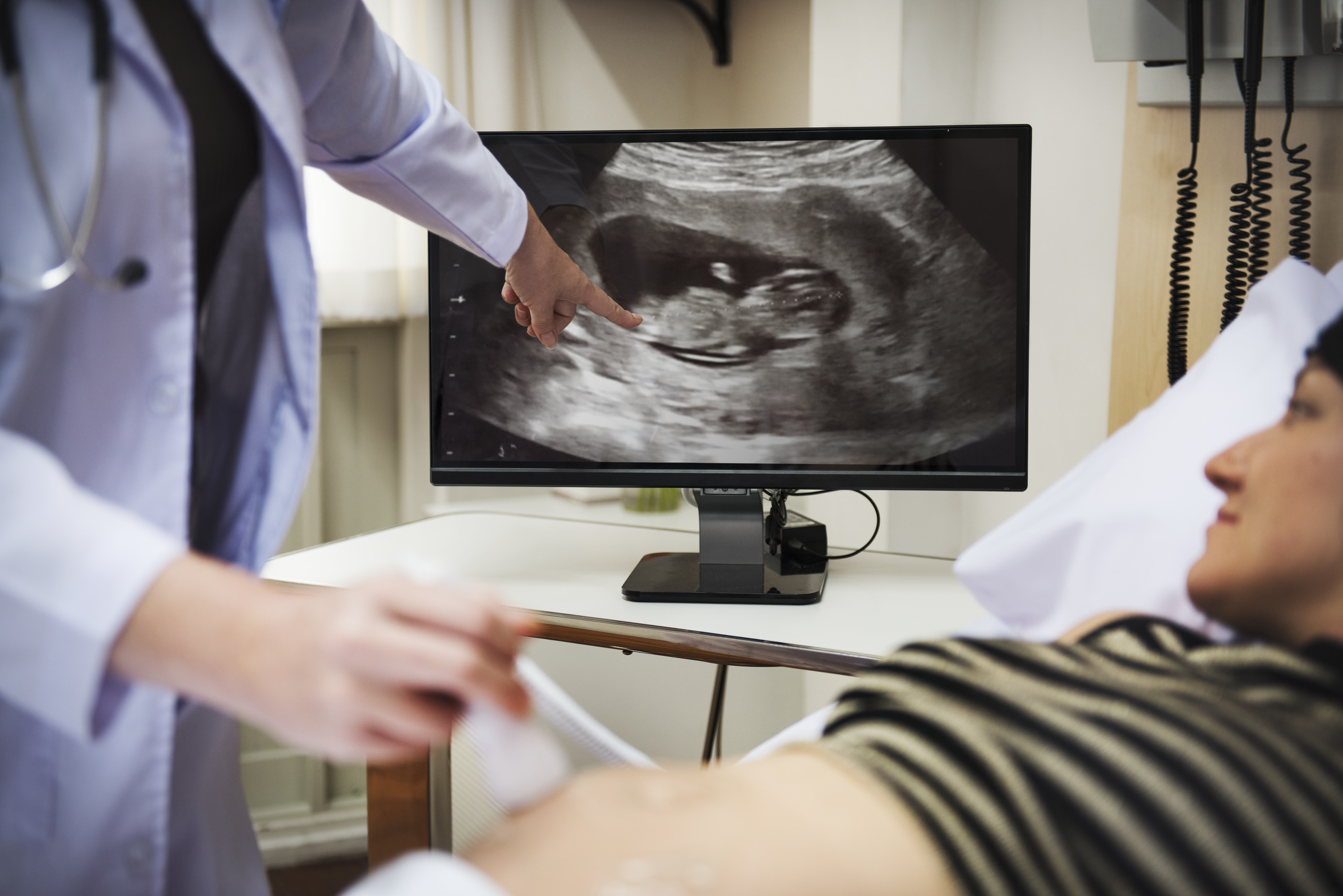
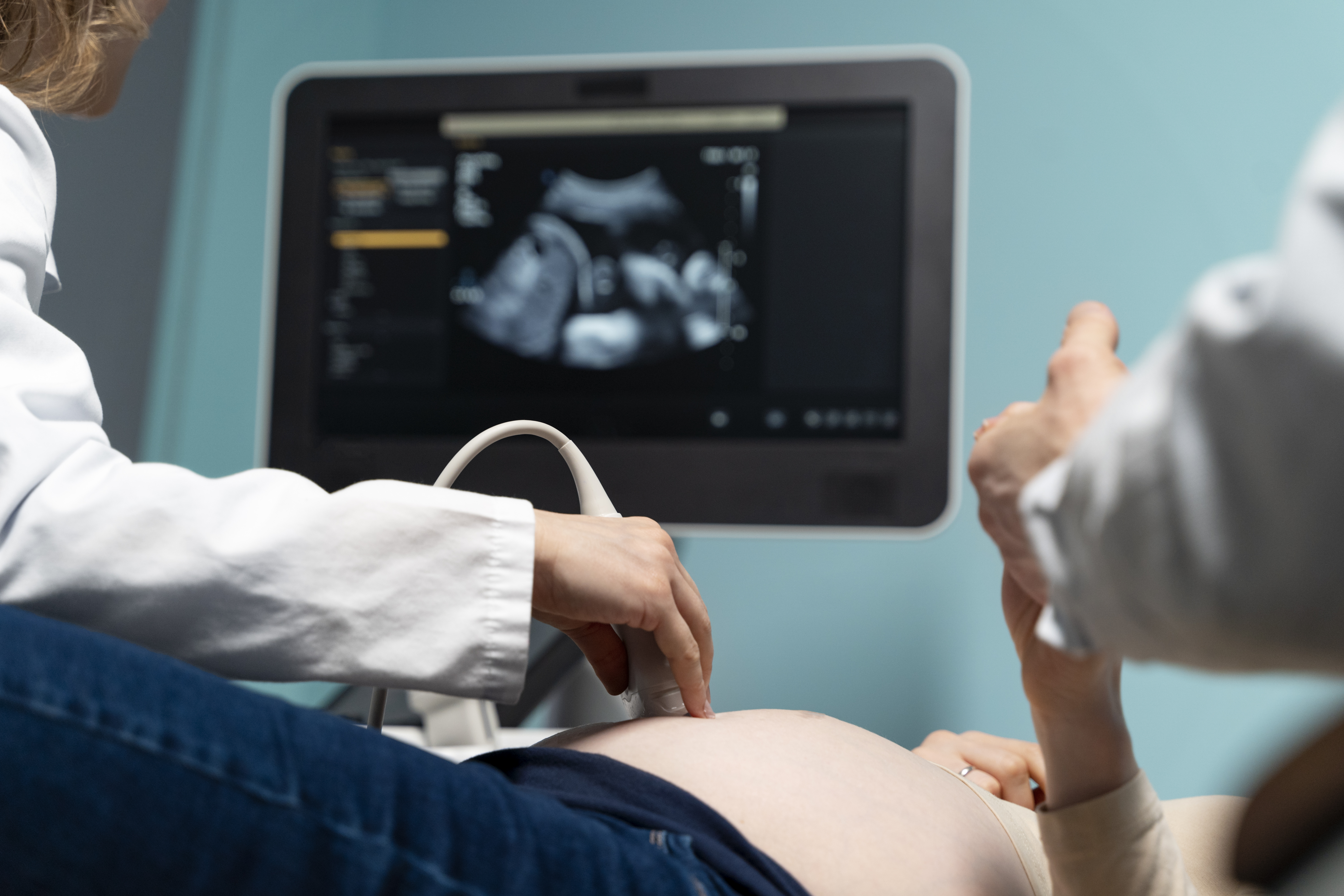
During the examination, ultrasound gel is applied to the abdomen and a transducer is moved over the skin.
You may feel some pressure while images are being obtained.
The level of pressure or discomfort can vary depending on:
- The position of the fetus
- The area being examined
- The amount of pressure needed to obtain images
- Your individual comfort
Tell the healthcare professional if you experience significant discomfort during the examination.
How Should You Prepare for a 3D/4D Obstetric Scan?
Preparation depends on the type and timing of the obstetric ultrasound.
Not every pregnancy scan requires the same preparation.
Before your appointment, ask the centre whether you should:
- Drink water before the scan
- Have a partially or fully filled bladder
- Avoid eating or drinking for a particular period
- Bring previous pregnancy scan reports
- Bring your doctor's prescription or referral
Do not assume that the preparation for one pregnancy scan is the same as another.
Bring Previous Reports
Previous ultrasound reports can provide useful clinical context for your healthcare team, particularly when follow-up imaging is being performed.
What Happens During a 3D/4D Obstetric Scan?
Understanding the process can help you feel prepared.
1. Patient Information
The healthcare professional may ask about:
- Gestational age
- Previous scans
- Medical history
- Pregnancy history
- Current concerns
- Doctor's referral
2. Positioning
You will usually lie comfortably on the examination bed.
3. Ultrasound Gel
A layer of ultrasound gel is applied to the abdomen.
4. Image Acquisition
The transducer is moved across the abdomen to obtain images.
5. 3D/4D Imaging
When suitable images can be obtained, the ultrasound system may reconstruct three-dimensional views and, with 4D imaging, display movement in real time.
6. Additional Measurements
Depending on the examination, the healthcare professional may obtain measurements and assess specific fetal or pregnancy-related structures.
Why Can 3D/4D Ultrasound Images Look Different From One Pregnancy to Another?
The quality and appearance of 3D/4D images can vary considerably between pregnancies.
Factors that can influence image quality include:
- Fetal position
- Gestational age
- Fetal movement
- Amount of amniotic fluid
- Placental location
- Maternal body characteristics
- Position of the fetus relative to the ultrasound probe
Therefore, a less clear 3D/4D image does not automatically indicate a problem with the pregnancy.
The diagnostic value of an obstetric ultrasound is not determined solely by how clear a 3D facial image appears.
What Is Doppler Ultrasound in Pregnancy?
Doppler ultrasound is a specialized ultrasound technique used to evaluate blood flow.
During pregnancy, Doppler techniques may be used in selected clinical situations to assess blood flow in areas such as the:
- Umbilical cord
- Placenta
- Fetal circulation
- Maternal blood vessels
Doppler is not required for every pregnancy scan.
Its use should be based on the clinical indication and performed appropriately by trained professionals. The AIUM recommends that Doppler techniques be used judiciously, particularly during early pregnancy.
Can a 3D/4D Scan Detect All Birth Defects?
No. No ultrasound examination can detect every fetal abnormality.
Ultrasound can help identify many anatomical findings, but some abnormalities may be difficult to detect depending on:
- Gestational age
- Fetal position
- Image quality
- Type of examination
- Nature of the abnormality
- Clinical circumstances
RadiologyInfo notes that obstetric ultrasound cannot identify all fetal abnormalities. Further testing may sometimes be required when there is a clinical or laboratory concern.
A normal ultrasound is reassuring, but it does not guarantee that every possible condition has been ruled out.
Is 3D/4D Ultrasound the Same as an Anomaly Scan?
No. They are related to pregnancy imaging but should not automatically be considered the same examination.
An anomaly scan, often performed during the second trimester, is a detailed ultrasound examination intended to assess fetal anatomy and look for certain structural abnormalities.
A 3D/4D scan refers primarily to the way ultrasound data is reconstructed and displayed.
3D or 4D imaging may sometimes be used as part of a specialized obstetric examination, but 3D/4D imaging does not replace a clinically appropriate detailed fetal anatomical examination.
The examination you need should be based on your obstetrician's recommendation.
3D/4D Ultrasound vs Standard Pregnancy Ultrasound
Both are ultrasound examinations, but they may serve different purposes.
| Standard Pregnancy Ultrasound | 3D/4D Ultrasound |
|---|---|
| Primarily provides conventional 2D diagnostic images | Provides 3D images and, with 4D, real-time movement |
| Used for routine pregnancy assessment and specific clinical questions | Can provide additional spatial and dynamic views when appropriate |
| Can assess fetal growth, position and other pregnancy findings | Can provide additional views of selected fetal structures |
| May be sufficient for many clinical assessments | Does not replace every standard or specialized obstetric examination |
The most appropriate examination is the one that answers the medical question your doctor needs addressed.
What Happens After a 3D/4D Obstetric Scan?
After the examination, the images and findings are reviewed as part of the diagnostic process.
The interpretation may be considered together with:
- Gestational age
- Previous ultrasound findings
- Maternal medical history
- Pregnancy history
- Clinical findings
- Laboratory results
- Other investigations
Your treating obstetrician or appropriate healthcare professional can explain what the findings mean for your pregnancy.
If additional testing or follow-up ultrasound is recommended, it does not necessarily mean that something is wrong. Follow-up may simply be needed to obtain additional views or monitor a finding over time.
Why Choose AVR Scan Centre for a 3D/4D Obstetric Scan in OMR?
When choosing a 3D/4D Obstetric Scan Centre in OMR, patients may want to consider the availability of the required pregnancy examination, location, preparation requirements and appointment process.
AVR Scan Centre provides 3D/4D Obstetric Scan services for pregnancy-related diagnostic imaging.
1. 3D/4D Obstetric Imaging
AVR Scan Centre offers 3D/4D obstetric imaging for patients whose doctors have recommended this type of pregnancy ultrasound.
2. OMR-Side Location
The Sholinganallur centre serves patients looking for pregnancy imaging in and around the OMR region.
3. Patient Preparation Guidance
Patients can contact the centre before the appointment to confirm the examination type and any preparation required.
4. Other Diagnostic Services
AVR Scan Centre also provides several diagnostic services, including:
- MRI Scan 1.5T
- MultiSlice 3D Spiral CT Scan
- Ultrasound
- X-Ray
- Computerized ECG
- Echocardiogram
- SonoMammography
- Treadmill Test
- EEG
- PFT
- Lab Services
- Preventive Master Health Checkup
- Digital Mammogram
- OPG
What Should You Check Before Booking a 3D/4D Obstetric Scan in OMR?
Before scheduling your pregnancy ultrasound, confirm:
- The exact type of pregnancy scan recommended
- Your current gestational age
- Whether the scan is routine, detailed or targeted
- Whether 3D/4D imaging is clinically indicated
- Whether Doppler assessment has been requested
- Whether any preparation is required
- Whether you need previous pregnancy reports
- Whether an appointment is required
Before You Visit AVR Scan Centre
Confirm the exact examination and preparation instructions with the centre before your appointment.
This is important because different pregnancy ultrasound examinations can have different requirements.
Frequently Asked Questions About 3D/4D Obstetric Scan in OMR
A 3D obstetric ultrasound creates three-dimensional images from ultrasound data, while 4D ultrasound adds real-time movement to those images.
AVR Scan Centre provides 3D/4D Obstetric Scan services for patients looking for pregnancy imaging in and around the OMR region. Contact the centre to confirm examination and appointment details.
Diagnostic ultrasound is considered safe when appropriately used for medical purposes. Professional guidance recommends prudent use and minimizing exposure while obtaining the necessary diagnostic information.
No. Ultrasound uses sound waves and does not use ionizing radiation.
There is no single best week for every pregnancy. Timing depends on the purpose of the examination, gestational age, fetal position and imaging conditions. Your obstetrician or healthcare professional can advise you about the appropriate timing.
No. A detailed anomaly scan focuses on fetal anatomy and screening for certain structural abnormalities, whereas 3D/4D refers to the way ultrasound images are generated and displayed. 3D/4D imaging does not automatically replace a detailed anatomical examination.
It may help visualize certain fetal structures and abnormalities, but no ultrasound examination can detect every fetal abnormality. Additional testing may sometimes be required.
Preparation varies depending on the examination and gestational age. Contact the centre before your appointment for the specific instructions.
Not necessarily. They provide different types of images. Standard 2D ultrasound remains important for many clinical assessments, while 3D/4D imaging can provide additional views in suitable situations.
Doppler ultrasound evaluates blood flow. During pregnancy, it may be used in selected situations to assess blood flow involving the placenta, umbilical cord or fetal circulation.
A 3D/4D ultrasound may produce views of the baby's face when fetal position and other imaging conditions allow. However, image quality varies and a clear facial image should not be the primary measure of the medical value of the examination.
Professional ultrasound organizations recommend that fetal ultrasound be performed for an appropriate medical purpose and discourage nonmedical or unnecessary ultrasound exposure.
The duration varies according to the examination, gestational age, fetal position and whether additional measurements or imaging are required. Ask the centre about the expected duration of your appointment.
Bring your doctor's prescription or referral and previous pregnancy scan reports when available. Follow any preparation instructions provided for your particular examination.
Depending on the gestational age, fetal position and local laws and regulations, ultrasound may sometimes visualize fetal sex. However, the purpose of prenatal ultrasound is medical assessment of the pregnancy, not sex selection or nonmedical fetal imaging.
Fetal position can affect image quality. Sometimes the desired view cannot be obtained clearly during a particular examination. Your healthcare professional can decide whether additional imaging or another assessment is appropriate.
Get Your 3D/4D Obstetric Scan at AVR Scan Centre, Sholinganallur
Pregnancy ultrasound is an important part of prenatal care. Understanding which examination your doctor has recommended, when it should be performed and how to prepare can help you arrive prepared for your appointment.
AVR Scan Centre provides 3D/4D Obstetric Scan services for patients looking for pregnancy imaging in and around OMR.
Before visiting, contact the centre to confirm:
- Your required pregnancy scan
- Appointment availability
- Preparation instructions
- Whether 3D/4D imaging is part of the recommended examination
- Whether you need to bring previous pregnancy reports
Contact AVR Scan Centre – Sholinganallur to enquire about the pregnancy ultrasound recommended by your doctor and confirm appointment availability.
AVR Scan Centre – Sholinganallur
First Floor, No. 278/1 & 278/2, Commercial Building,
Ramanyam Pushkar 2, Kalaignar Karunanidhi Salai,
Sholinganallur – 600119.
