
OPG X-Ray in OMR
Panoramic Dental Imaging at AVR Scan Centre
If your dentist has recommended an OPG X-Ray in OMR, you may want to understand what an OPG shows, why it is recommended, whether it is painful, how to prepare and how it differs from a regular dental X-ray.
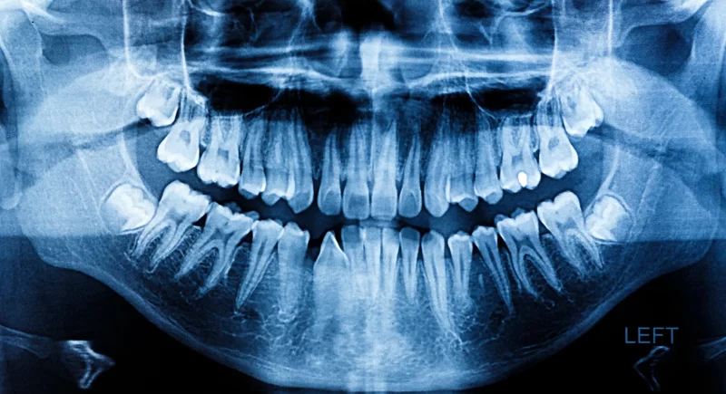
OPG stands for Orthopantomogram. It is a panoramic dental X-ray that captures a broad view of the teeth, upper and lower jaws and surrounding structures in a single image.
It can be useful for dental assessment, treatment planning and evaluating certain jaw and tooth-related conditions.
Unlike small intraoral dental X-rays, where the detector is positioned inside the mouth, an OPG uses equipment that rotates around the patient's head to create a panoramic image.
AVR Scan Centre provides OPG services for patients looking for dental and jaw imaging in and around the OMR region.
The exact type of dental imaging you need should be determined by your dentist or oral healthcare professional based on your symptoms, examination and treatment requirements.
What Is an OPG X-Ray?
An OPG, or Orthopantomogram, is a panoramic dental X-ray that produces a broad two-dimensional image of the teeth, upper and lower jaws and surrounding structures.
It provides a wider field of view than many individual intraoral dental X-rays and can help a dentist or oral healthcare professional assess:
- Teeth
- Upper jaw
- Lower jaw
- Developing or impacted teeth
- Wisdom teeth
- Jaw bone
- Selected jaw abnormalities
- Surrounding structures visible on the panoramic image
An OPG is a 2D panoramic examination, not a 3D scan.
If more detailed three-dimensional information is required, your dentist may recommend another examination, such as dental cone-beam CT, depending on the clinical question.
OPG X-Ray in OMR – Where Can You Get an OPG Scan?
If you are searching for an OPG X-Ray in OMR, AVR Scan Centre provides OPG diagnostic imaging services for patients in and around the OMR region.
The Sholinganallur centre serves patients looking for diagnostic services around OMR and nearby localities.
Patients travelling from Padur, Navalur, Siruseri, Kelambakkam, Thiruporur and surrounding areas can contact AVR Scan Centre before visiting to confirm OPG availability and appointment requirements.
Before your appointment, confirm:
- OPG availability
- Appointment requirements
- Whether any specific preparation is required
- Whether you need to bring previous dental X-rays
- Whether your dentist has requested any additional imaging
AVR Scan Centre – Sholinganallur
First Floor, No. 278/1 & 278/2, Commercial Building, Ramanyam Pushkar 2, Kalaignar Karunanidhi Salai, Sholinganallur – 600119.
Why Might a Dentist Recommend an OPG?
An OPG may be recommended when a broad panoramic view of the teeth and jaws is useful for diagnosis or treatment planning.
Depending on the clinical situation, an OPG may be used for:
- Evaluating impacted wisdom teeth
- Assessing tooth positioning
- Planning dental extractions
- Planning orthodontic treatment
- Assessing dental implants
- Evaluating dentures
- Examining the jaw bones
- Assessing selected jaw abnormalities
- Evaluating certain temporomandibular joint-related concerns
- Assessing some dental and oral conditions
The exact reason for an OPG depends on your dental examination and treatment plan.
What Can an OPG X-Ray Show?
An OPG provides a broad overview of the teeth and jaw structures that can be included in the panoramic image.
Teeth
An OPG can provide information about the position and development of teeth across the upper and lower jaws.
Wisdom Teeth
Panoramic imaging can be useful for assessing the position and development of wisdom teeth, including teeth that have not fully erupted.
Impacted Teeth
An impacted tooth is one that has not erupted normally. An OPG can help show the position of an unerupted or impacted tooth.
Jaw Bone
The panoramic image can provide an overview of the upper and lower jaw bones and may show selected abnormalities.
Dental Treatment Planning
An OPG may be used as part of treatment planning for:
- Braces
- Extractions
- Dentures
- Dental implants
- Selected oral and maxillofacial procedures
What Conditions Can an OPG Help Evaluate?
An OPG can help healthcare professionals identify or assess certain dental and jaw-related abnormalities, but it is not the best test for every dental condition.
Depending on the clinical indication, panoramic imaging may contribute to the evaluation of:
- Advanced periodontal disease
- Impacted teeth
- Jaw cysts
- Certain jaw tumours or other abnormalities
- Temporomandibular joint disorders
- Certain sinus-related findings
- Dental and jaw development
- Tooth positioning
The OPG findings need to be interpreted together with the clinical examination.
How Does an OPG X-Ray Work?
During an OPG examination, the X-ray tube and detector move around the patient's head to capture a panoramic image of the mouth and jaws.
The patient stands or sits in the OPG unit while the head is positioned carefully.
A bite block or positioning guide may be used to help align the teeth and jaws correctly. The rotating equipment then moves around the head while the image is acquired.
The resulting image provides a broad panoramic view of the dental and jaw structures.
What Happens During an OPG Scan?
An OPG is a quick and non-invasive dental X-ray examination.
1. Patient Positioning
You will be positioned in the centre of the panoramic X-ray unit. Your head needs to be aligned correctly to help obtain a clear image.
2. Removing Metal Objects
You may be asked to remove:
- Earrings
- Neck jewellery
- Spectacles
- Other metal objects that could interfere with the image
3. Bite Positioning
You may be asked to place your teeth on or around a positioning device to help maintain the correct alignment.
4. Staying Still
You will need to remain very still while the equipment rotates around your head.
5. Image Acquisition
The rotating arm captures the panoramic image. The actual image acquisition is usually very quick; RadiologyInfo notes that it can typically take around 12–20 seconds.
6. Completion
Once the image has been captured, you can leave the examination area unless additional imaging has been requested.
How Should You Prepare for an OPG X-Ray?
An OPG generally requires little or no special preparation.
Remove Jewellery and Metal Objects
Remove metal objects around the head and neck that may interfere with the X-ray image.
Remove Spectacles
You may be asked to remove your glasses during image acquisition.
Tell the Imaging Team About Pregnancy
Inform the imaging professional if you are pregnant or may be pregnant. Because an OPG uses X-rays, the team can determine whether the examination is appropriate and apply appropriate radiation-safety measures.
Bring Previous Dental Images
Previous dental X-rays can be useful when comparison is needed.
Does an OPG X-Ray Use Radiation?
Yes. An OPG uses a small amount of ionizing radiation to create the panoramic dental image.
As with other X-ray examinations, radiation protection principles are used to minimize unnecessary exposure.
RadiologyInfo lists an approximate effective radiation dose for a panoramic X-ray of about 0.025 mSv, although actual dose varies with equipment, technique and patient factors.
The FDA and American Dental Association guidance emphasizes that dental radiographs should be selected based on individual clinical need and performed using radiation-minimization principles.
Is an OPG X-Ray Safe?
An OPG is a commonly used dental X-ray examination, and the radiation exposure is low, but it should be performed when clinically appropriate.
The decision to perform a dental X-ray should consider:
- Your dental symptoms
- Clinical findings
- Treatment requirements
- Previous imaging
- Whether the information can be obtained without another radiograph
Dental imaging guidelines emphasize individualized selection of radiographs to avoid unnecessary radiation exposure.
Is an OPG X-Ray Painful?
No. An OPG examination is generally painless and non-invasive.
There are no needles or surgical procedures involved.
You simply need to remain correctly positioned while the panoramic equipment moves around your head.
Some patients may find keeping the bite position or remaining still slightly uncomfortable, but the image acquisition itself is very brief.
OPG vs Normal Dental X-Ray – What Is the Difference?
| OPG / Panoramic X-Ray | Intraoral Dental X-Ray |
|---|---|
| Provides a broad view of the teeth and jaws | Provides detailed images of a smaller area |
| Detector remains outside the mouth | Detector is placed inside the mouth |
| Useful for panoramic assessment | Useful for detailed evaluation of individual teeth and nearby structures |
| Can show tooth positioning and jaw structures | Often provides greater detail for specific teeth and roots |
| Useful for certain treatment-planning situations | Useful for detailed dental assessment |
Panoramic imaging covers a wider area, while intraoral radiographs can provide more precise detail for particular teeth and dental structures.
Is an OPG Better Than a Dental X-Ray?
Neither is universally better. Your dentist chooses the appropriate X-ray based on what needs to be examined.
OPG vs Dental CBCT – What Is the Difference?
An OPG produces a two-dimensional panoramic image, while dental CBCT creates three-dimensional images of selected dental and maxillofacial structures.
| OPG | Dental CBCT |
|---|---|
| 2D panoramic image | 3D volumetric imaging |
| Broad overview of teeth and jaws | Detailed 3D visualization of selected regions |
| Useful for many routine dental assessments | Useful when three-dimensional information is required |
| Lower radiation dose than many CBCT examinations | Radiation dose varies by CBCT system and protocol |
| Useful for initial assessment in many situations | Selected for more detailed treatment planning or specific clinical questions |
CBCT should not automatically be considered better than OPG. The appropriate examination depends on the clinical question and the amount of information your dentist needs.
Can an OPG Detect Wisdom Tooth Problems?
Yes. An OPG can provide a broad view of wisdom teeth and their position.
It can help show whether wisdom teeth are:
- Erupted
- Partially erupted
- Unerupted
- Impacted
- Positioned unusually relative to surrounding teeth
Panoramic imaging is commonly used when assessing the development and position of wisdom teeth.
If more detailed information is needed for treatment planning, your dentist may recommend additional imaging.
Can an OPG Be Used for Braces and Orthodontic Treatment?
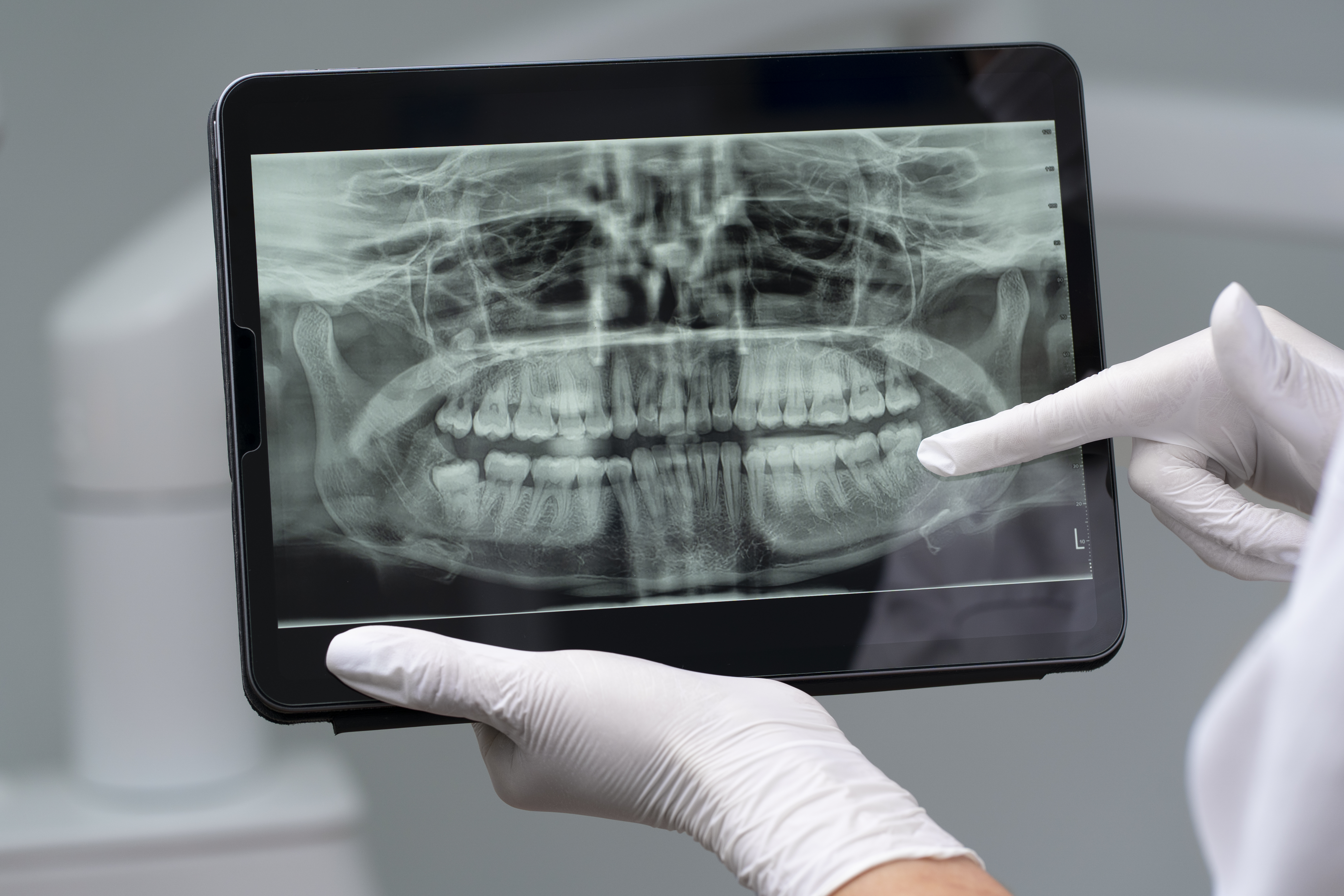
Yes. Panoramic imaging can be useful as part of orthodontic assessment and treatment planning.
It can provide a broad view of:
- Tooth position
- Developing teeth
- Eruption status
- Root development
- Jaw structures
The orthodontist may combine OPG findings with other examinations and imaging depending on the treatment plan.
Can an OPG Be Used Before Dental Implants?
An OPG may be used as part of an initial dental implant assessment, but it may not provide all the three-dimensional information needed for implant planning.
An OPG can give a broad overview of the jaw and teeth.
If detailed measurements of bone width, bone height or the relationship between anatomical structures are required, a dentist may recommend a dental CBCT examination.
The appropriate imaging should be selected by the dental professional planning the treatment.
Can an OPG Detect Jaw Problems?
An OPG can show certain abnormalities involving the teeth and jaw bones, but it does not provide detailed information about every jaw or soft-tissue condition.
Depending on the clinical situation, it may help identify or evaluate:
- Jaw bone abnormalities
- Impacted teeth
- Certain cystic lesions
- Selected tumours or other abnormalities
- Some TMJ-related findings
- Tooth and jaw development
Because panoramic imaging has limitations, additional imaging such as CBCT or other examinations may be required for some conditions.
Can an OPG Detect Oral Cancer?
An OPG may show some abnormalities involving the jaw or surrounding structures, but it is not a standalone test for diagnosing oral cancer.
If a dentist or doctor finds a suspicious oral lesion, additional clinical evaluation and, when appropriate, tissue biopsy may be required.
An OPG should therefore be viewed as one part of a diagnostic assessment rather than a complete oral-cancer screening test.
What Are the Limitations of an OPG X-Ray?
An OPG provides a broad overview, but it does not provide highly detailed information about every tooth, soft tissue or jaw structure.
Limitations can include:
- Less detail than some intraoral dental X-rays for individual teeth
- Two-dimensional representation of three-dimensional anatomy
- Overlapping structures
- Image distortion
- Potential blurring
- Limited information about soft tissues
RadiologyInfo notes that panoramic X-rays are generally used as an initial evaluation of teeth and bones, and additional imaging may be required when more precise information is needed.
What Happens After an OPG X-Ray?
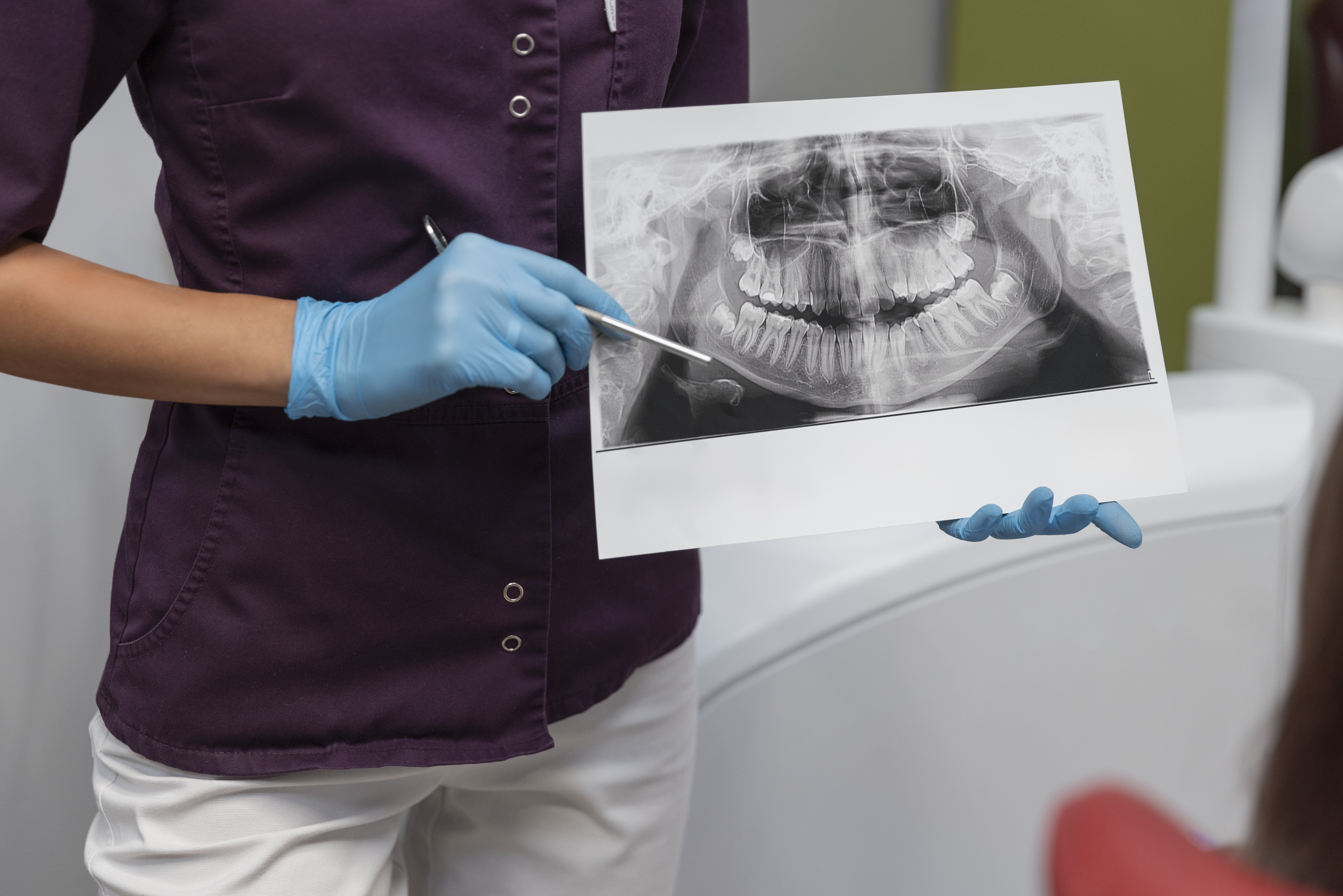
Once the panoramic image has been captured, it can be reviewed as part of your dental assessment.
Your dentist may consider the OPG alongside:
- Clinical examination
- Symptoms
- Dental history
- Previous X-rays
- Treatment requirements
- Other diagnostic findings
If additional detail is required, your dentist may recommend another examination.
Do not attempt to diagnose a dental condition from the panoramic image alone.
Why Choose AVR Scan Centre for an OPG in OMR?
When looking for an OPG X-Ray Centre in OMR, patients may want to consider location, availability of the required dental X-ray, appointment requirements and access to related diagnostic imaging.
AVR Scan Centre provides OPG services for patients seeking dental and jaw imaging in and around the OMR region.
1. OPG Diagnostic Service
AVR Scan Centre provides OPG imaging as part of its diagnostic services.
2. OMR-Side Location
The Sholinganallur centre serves patients looking for diagnostic services around the OMR area.
3. Patient Preparation Guidance
Patients can contact the centre before their appointment to confirm the OPG examination process and any instructions before visiting.
AVR Scan Centre also provides:
- MRI Scan 1.5T
- MultiSlice 3D Spiral CT Scan
- Ultrasound
- X-Ray
- 3D/4D Obstetrics
- Computerized ECG
- Echocardiogram
- SonoMammography
- Treadmill Test
- EEG
- PFT
- Lab Services
- Preventive Master Health Checkup
- Digital Mammogram
What Should You Check Before Booking an OPG X-Ray in OMR?
Before scheduling your OPG, confirm:
- Why your dentist has recommended the examination
- Whether an OPG is sufficient for the clinical question
- Whether you need additional dental imaging
- Whether you have previous dental X-rays
- Whether you are pregnant or may be pregnant
- Whether an appointment is required
Before You Visit AVR Scan Centre
Bring your dentist's prescription or referral where applicable and previous dental imaging reports if available.
Tell the imaging professional if you are pregnant or may be pregnant before the X-ray examination.
Frequently Asked Questions About OPG X-Ray in OMR
OPG stands for Orthopantomogram. It is a panoramic dental X-ray that creates a broad 2D image of the teeth, upper and lower jaws and surrounding structures.
AVR Scan Centre provides OPG services for patients looking for dental and jaw imaging in and around OMR. Contact the centre to confirm availability and appointment requirements.
Yes. An OPG uses a small amount of ionizing X-ray radiation to create the panoramic dental image.
No. OPG is generally painless and non-invasive. You simply need to remain properly positioned while the panoramic image is acquired.
No special preparation is generally required for a panoramic dental X-ray.
Yes. An OPG provides a broad view of wisdom teeth and can help assess their position and development.
Yes. Panoramic imaging can show unerupted and impacted teeth and their position relative to surrounding structures.
Yes. An OPG can be useful in orthodontic assessment and treatment planning, depending on the clinical requirement.
It can be useful as part of an initial assessment, but additional 3D imaging such as CBCT may be needed when detailed implant planning is required.
OPG produces a 2D panoramic image, while CBCT provides 3D images of selected dental and maxillofacial structures.
OPG provides a broad view of the teeth and jaws, while intraoral X-rays generally provide more detailed images of smaller areas, such as individual teeth.
An OPG may show some dental abnormalities, but small cavities are often better evaluated with intraoral dental X-rays and clinical examination. Your dentist will select the appropriate imaging.
It can show certain jaw bone abnormalities and other findings, but additional imaging may be necessary for more detailed assessment.
Tell your dentist and the imaging team if you are pregnant or may be pregnant. They can determine whether the examination is necessary and apply appropriate radiation-safety precautions.
The image acquisition itself is very brief, typically around 12–20 seconds, although positioning and the overall appointment take longer.
Bringing previous dental X-rays can be useful when comparison is required.
Your dentist will interpret the findings along with your clinical examination and symptoms. Additional dental X-rays, CBCT, other imaging or treatment may be recommended depending on the finding.
No. OPG provides a broad panoramic overview but does not provide the same level of detail as every intraoral dental X-ray. The appropriate imaging depends on what your dentist needs to assess.
Get Your OPG X-Ray at AVR Scan Centre, Sholinganallur
An OPG can provide a useful panoramic view of the teeth and jaws when a dentist needs broader dental imaging for diagnosis or treatment planning.
Understanding why your dentist has recommended an OPG and whether additional dental imaging is required can help you prepare for the examination.
AVR Scan Centre provides OPG services for patients looking for dental and jaw imaging in and around OMR.
Before visiting, contact the centre to confirm:
- OPG availability
- Appointment requirements
- Any specific instructions
- Whether additional imaging has been requested
Contact AVR Scan Centre – Sholinganallur to enquire about the OPG examination recommended by your dentist and confirm appointment availability.
AVR Scan Centre – Sholinganallur
First Floor, No. 278/1 & 278/2, Commercial Building,
Ramanyam Pushkar 2, Kalaignar Karunanidhi Salai,
Sholinganallur – 600119.
