
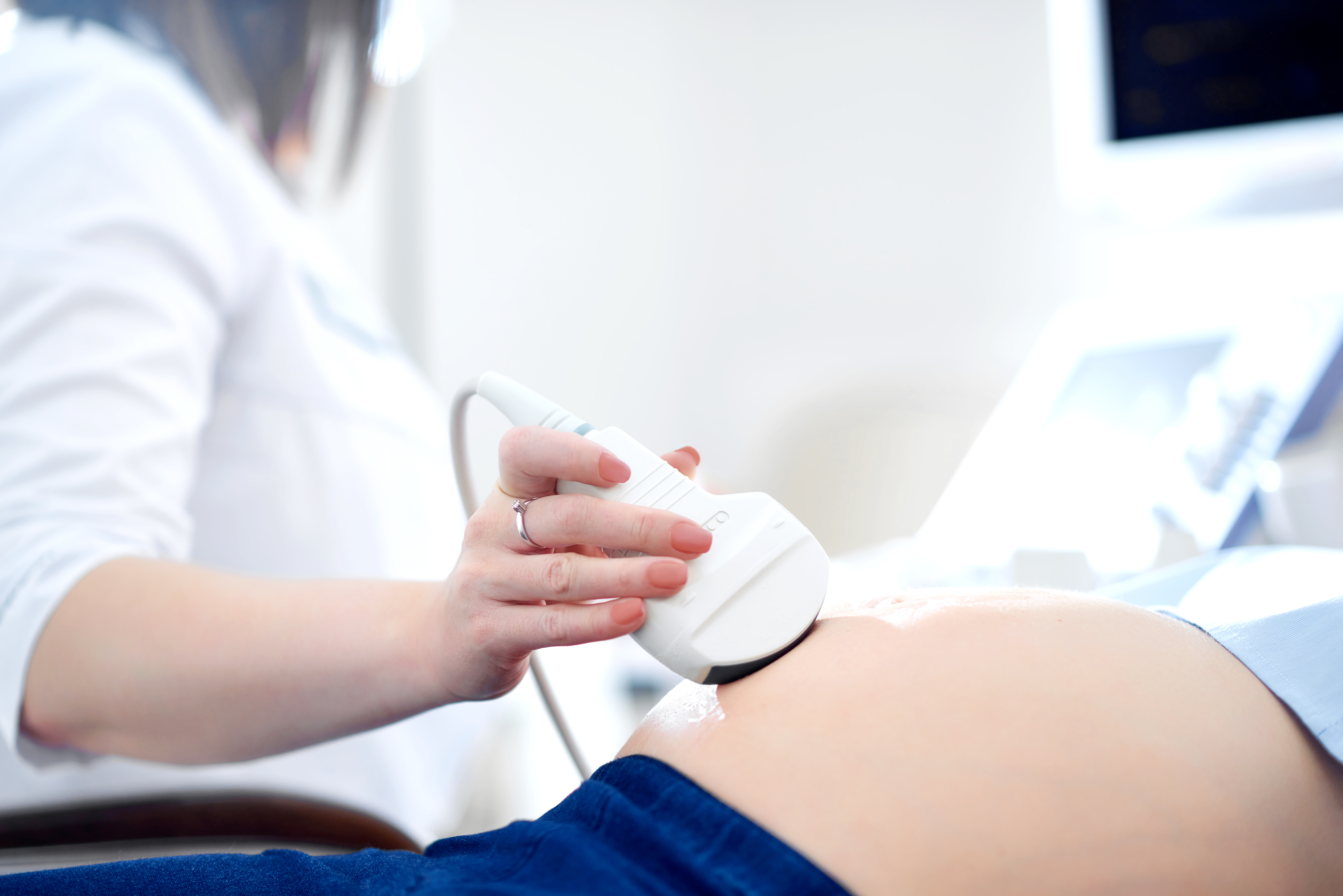
Ultrasound Scan in OMR
Diagnostic Ultrasound at AVR Scan Centre
If your doctor has recommended an Ultrasound Scan in OMR, you may want to know what the examination can show, whether you need to prepare, whether ultrasound is safe, and what happens during the scan.
Ultrasound imaging, also called sonography, uses high-frequency sound waves to create real-time images of structures inside the body. Unlike X-rays and CT scans, ultrasound does not use ionizing radiation. It can be useful for examining many soft tissues and internal organs and is also widely used during pregnancy when clinically indicated.
AVR Scan Centre provides Ultrasound services for patients looking for diagnostic imaging in and around OMR.
The exact type of ultrasound you need should be based on your doctor's recommendation because preparation and the scanning method can differ according to the body part being examined.
What Is an Ultrasound Scan?
An ultrasound scan is a non-invasive diagnostic imaging examination that uses high-frequency sound waves to produce images of structures inside the body.
A small device called a transducer is moved over the skin, usually with ultrasound gel between the transducer and the skin. The sound waves travel into the body and return as echoes. The ultrasound system processes these echoes to create images.
Ultrasound can provide real-time imaging and may be used to examine:
- Abdominal organs
- Pelvic structures
- Urinary system
- Thyroid and other superficial structures
- Soft tissues
- Blood flow through vessels
- Pregnancy and fetal development when medically indicated
The type of ultrasound depends on the clinical question your doctor needs to answer.
Ultrasound Scan in OMR – Where Can You Get an Ultrasound?
If you are searching for an Ultrasound Scan in OMR, AVR Scan Centre offers ultrasound diagnostic services for patients in and around the OMR region.
The Sholinganallur centre is located in the OMR area, making it a convenient diagnostic imaging option for patients from nearby localities.
Patients travelling from Padur, Navalur, Siruseri, Kelambakkam, Thiruporur and surrounding areas can contact AVR Scan Centre before visiting to confirm the availability of the ultrasound examination recommended by their doctor.
Before your appointment, confirm:
- The exact type of ultrasound required
- Whether fasting is necessary
- Whether you need to drink water before the examination
- Appointment requirements
- Any specific preparation instructions
Because different ultrasound examinations have different preparation requirements, it is better to confirm your instructions before arriving.
Why Might a Doctor Recommend an Ultrasound Scan?
A doctor may recommend an ultrasound when real-time imaging of organs, soft tissues, fluid-filled structures or blood flow can help evaluate a medical concern.
The decision may depend on:
- Your symptoms
- Medical history
- Physical examination
- Previous test results
- Body area requiring evaluation
- Suspected condition
- Need for follow-up imaging
Ultrasound may be useful for evaluating selected conditions involving the abdomen, pelvis, urinary system, thyroid, soft tissues, blood vessels and pregnancy.
Your doctor will determine whether ultrasound is appropriate for your particular situation.
What Areas of the Body Can an Ultrasound Scan Examine?
Ultrasound can be used for different parts of the body depending on the examination requested.
Abdominal Ultrasound
An abdominal ultrasound can provide images of abdominal organs such as the:
- Liver
- Gallbladder
- Kidneys
- Pancreas
- Spleen
- Major abdominal blood vessels
The exact organs examined depend on the prescribed examination.
Pelvic Ultrasound
Pelvic ultrasound can be used to examine structures within the pelvis.
Depending on the patient and clinical requirement, different approaches may be used, including abdominal or other specialized pelvic ultrasound techniques.
Urinary System Ultrasound
Ultrasound may be used to examine structures such as the:
- Kidneys
- Urinary bladder
- Related urinary structures
Thyroid and Neck Ultrasound
Ultrasound can provide detailed images of superficial structures such as the thyroid and selected tissues in the neck.
Soft-Tissue Ultrasound
Ultrasound may be used to evaluate selected superficial soft tissues, depending on the clinical concern.
Breast Ultrasound
Breast ultrasound may be recommended in selected situations to evaluate breast tissue and specific areas of concern.
It may be used as a complementary examination depending on the clinical situation and other breast imaging findings.
Vascular and Doppler Ultrasound
Doppler ultrasound can be used to assess blood flow through blood vessels. Ultrasound can provide real-time information about blood flow and may be used for selected vascular examinations.
What Is Doppler Ultrasound?
Doppler ultrasound is an ultrasound technique used to evaluate blood flow through blood vessels.
It can provide information about:
- Direction of blood flow
- Movement of blood
- Blood-flow patterns
- Selected abnormalities affecting blood vessels
Doppler techniques may be incorporated into an ultrasound examination when clinically appropriate.
The specific type of Doppler examination depends on the body part and the clinical question.
What Is an Ultrasound Scan Used For?
Ultrasound has a wide range of diagnostic applications.
Depending on the type of examination, it may help evaluate:
- Abdominal pain
- Pelvic symptoms
- Certain urinary symptoms
- Selected soft-tissue abnormalities
- Thyroid abnormalities
- Fluid-filled structures
- Certain vascular conditions
- Pregnancy-related findings
Ultrasound can also be used to guide some minimally invasive procedures, such as selected needle biopsies or fluid aspiration, when clinically appropriate.
An ultrasound scan does not provide a diagnosis by itself. The images and findings need to be interpreted in the context of your symptoms, medical history and other clinical information.
How Does an Ultrasound Scan Work?
Ultrasound uses sound waves rather than X-rays to create images.
During a typical examination:
- Ultrasound gel is applied to the skin.
- The transducer is placed over the examination area.
- Sound waves travel into the body.
- The returning echoes are detected by the transducer.
- Computer processing converts the information into images.
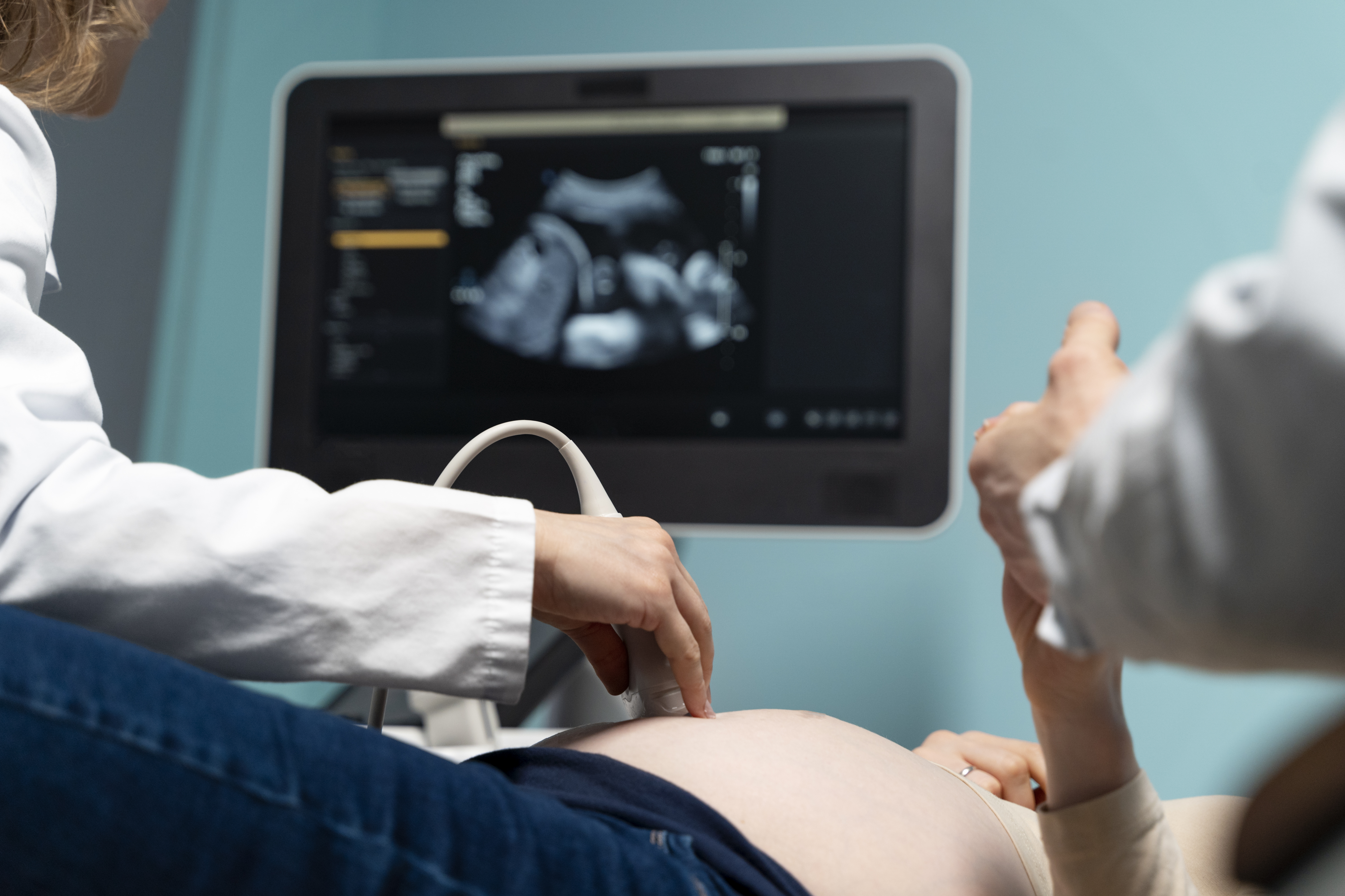
Because ultrasound produces images in real time, the examiner can observe certain movements and blood-flow patterns during the examination.
What Happens During an Ultrasound Scan?
An ultrasound examination is generally straightforward and does not usually require invasive procedures.
1. Preparation
Your preparation depends on the body part being examined.
2. Positioning
You will be asked to lie or sit in a position that allows the area to be examined properly.
3. Applying Ultrasound Gel
A small amount of gel is applied to the skin. The gel helps the transducer move smoothly and allows sound waves to travel effectively into the body.
4. Scanning
The sonographer or appropriately trained healthcare professional moves the transducer across the area being examined.
You may be asked to:
- Change position
- Take a breath
- Hold your breath briefly
- Drink water before the scan
- Have a full bladder for certain examinations
The instructions depend on the type of ultrasound.
5. Image Review
Images are obtained during the examination and form part of the diagnostic assessment.
How Should You Prepare for an Ultrasound Scan?
Ultrasound preparation depends on the type of examination. Some ultrasound scans require little or no preparation, while others may require fasting or a full bladder.
For Abdominal Ultrasound
You may be given instructions about fasting or food intake before the examination, depending on the specific abdominal scan.
For Pelvic Ultrasound
You may be asked to drink water before the examination so that your bladder is adequately filled, depending on the examination protocol.
For Other Ultrasound Examinations
Some examinations may require no special preparation.
Before Your Appointment
Contact the centre and confirm the specific preparation instructions for your ultrasound rather than following generic advice found online.
Is an Ultrasound Scan Safe?
Diagnostic ultrasound is considered safe when appropriately used and does not use ionizing radiation.
This is one reason ultrasound is widely used in different patient groups and during pregnancy when medically indicated.
However, ultrasound should still be used appropriately for a clinical purpose rather than performed unnecessarily.
During pregnancy, professional guidance recommends prudent use by appropriately trained healthcare professionals and keeping exposure as low as reasonably achievable while obtaining the required diagnostic information.
Does an Ultrasound Scan Use Radiation?
No. Ultrasound does not use ionizing radiation.
Ultrasound uses high-frequency sound waves to create images.
This makes it different from:
- X-Ray → Uses X-rays
- CT Scan → Uses X-rays
- MRI → Uses magnetic fields and radio waves
Because ultrasound does not use ionizing radiation, it is commonly used for examinations where repeated imaging may be needed and is also widely used during pregnancy when clinically appropriate.
Is an Ultrasound Scan Painful?
Most ultrasound examinations are non-invasive and are usually painless.
You may feel some pressure as the transducer moves over the skin, particularly when the examiner needs to obtain images from a particular area.
Some examinations may cause temporary discomfort depending on the body part being examined or the underlying condition.
Tell the healthcare professional if you experience significant discomfort during the examination.
Is Ultrasound Used During Pregnancy?
Yes. Ultrasound is widely used during pregnancy to assess the developing fetus and pregnancy-related structures when medically indicated.
What Are the Benefits of Pregnancy Ultrasound?
- Fetal development
- Fetal anatomy
- Pregnancy location
- Fetal growth
- Placental position
- Amniotic fluid
- Other pregnancy-related findings
The type and timing of an obstetric ultrasound depends on the pregnancy and the clinical reason for the examination.
Important
Ultrasound during pregnancy should be performed for an appropriate medical purpose by appropriately trained professionals. Non-medical fetal scanning purely for keepsake images is discouraged by the American Institute of Ultrasound in Medicine.
What Is the Difference Between Ultrasound and Doppler Ultrasound?
Both examinations use ultrasound technology, but they provide different types of information.
| Ultrasound | Doppler Ultrasound |
|---|---|
| Produces images of structures and organs | Evaluates blood flow |
| Used for many organs and soft tissues | Used particularly for blood vessels and blood-flow assessment |
| Can provide real-time imaging | Adds information about movement of blood |
| Examination depends on the body part | Used when vascular or blood-flow information is clinically required |
A Doppler study may be included as part of an ultrasound examination when clinically appropriate.
Ultrasound vs CT Scan – What Is the Difference?
Ultrasound and CT use different technologies and are selected for different clinical purposes.
| Ultrasound | CT Scan |
|---|---|
| Uses sound waves | Uses X-rays |
| Does not use ionizing radiation | Uses ionizing radiation |
| Provides real-time imaging | Provides cross-sectional imaging |
| Useful for many soft tissues and fluid-filled structures | Useful for many anatomical regions and clinical situations |
| Can assess blood flow using Doppler techniques | Can provide detailed cross-sectional and reconstructed images |
Is Ultrasound Better Than CT?
Neither ultrasound nor CT is universally better.
The appropriate examination depends on:
- Symptoms
- Body part
- Suspected condition
- Medical history
- Clinical question
- Information required by the doctor
Your doctor may recommend ultrasound, CT, MRI, X-ray or another examination depending on your individual situation.
Can Ultrasound Detect Everything?
No. Ultrasound is a valuable diagnostic tool, but it has limitations.
Ultrasound does not provide equally useful images of every body structure.
For example, sound waves can be affected by:
- Air or gas
- Bone
- Body size and tissue depth
- Patient movement
- The location of the structure being examined
Ultrasound is generally less suitable for imaging air-filled lungs and cannot pass through bone to visualize structures beyond it in the same way as CT or MRI.
If ultrasound does not answer the clinical question, your doctor may recommend another imaging examination.

What Happens After an Ultrasound Scan?
After the examination, the images and findings are reviewed as part of the diagnostic process.
The interpretation may be considered together with:
- Your symptoms
- Medical history
- Clinical examination
- Previous test results
- Other imaging studies
Your doctor can explain the findings and advise whether further investigation or follow-up is required.
In some situations, another imaging examination may be recommended if additional information is needed.
Do not interpret ultrasound images on your own or assume that an image automatically confirms a particular condition.
Why Choose AVR Scan Centre for an Ultrasound Scan in OMR?
When looking for an Ultrasound Scan in OMR, patients may want to consider the centre's location, availability of the required examination, preparation instructions and appointment process.
AVR Scan Centre provides ultrasound diagnostic services for patients seeking imaging in and around the OMR region.
1. Ultrasound Diagnostic Service
Ultrasound provides non-invasive imaging for a range of clinical requirements.
2. OMR-Side Location
AVR Scan Centre's Sholinganallur centre serves patients looking for diagnostic imaging around the OMR area.
3. Patient Preparation Information
Patients can contact the centre to confirm the exact ultrasound examination and any preparation required before the appointment.
4. Multiple Diagnostic Services
AVR Scan Centre also provides diagnostic services including:
- MRI Scan 1.5T
- MultiSlice 3D Spiral CT Scan
- X-Ray
- 3D/4D Obstetrics
- Computerized ECG
- Echocardiogram
- SonoMammography
- Treadmill Test
- EEG
- PFT
- Lab Services
- Preventive Master Health Checkup
- Digital Mammogram
- OPG
What Should You Check Before Booking an Ultrasound Scan in OMR?
Before scheduling your ultrasound, confirm:
- Which body part needs to be examined
- The specific type of ultrasound recommended
- Whether fasting is required
- Whether you need a full bladder
- Whether Doppler imaging is required
- Whether you need to bring previous reports
- Whether an appointment is required
- Any special preparation instructions
Before You Visit AVR Scan Centre
Confirm the exact ultrasound examination and preparation instructions with the centre before your appointment. This is particularly important because preparation varies between abdominal, pelvic, obstetric, vascular and other ultrasound examinations.
Frequently Asked Questions About Ultrasound Scan in OMR
An ultrasound scan is a non-invasive diagnostic imaging examination that uses high-frequency sound waves to create images of structures inside the body.
AVR Scan Centre provides ultrasound diagnostic services for patients looking for imaging around the OMR region. Contact the centre to confirm the required examination and appointment details.
No. Ultrasound uses sound waves and does not use ionizing radiation.
Most ultrasound examinations are non-invasive and generally painless, although some patients may experience temporary pressure or discomfort during certain examinations.
It depends on the type of ultrasound. Some examinations may require fasting, while others require little or no preparation. Follow the instructions for your specific examination.
Some pelvic and urinary ultrasound examinations may require a full bladder. The preparation depends on the examination, so follow the instructions provided for your appointment.
Doppler ultrasound is an ultrasound technique used to assess blood flow through blood vessels.
Diagnostic ultrasound is widely used during pregnancy when medically indicated. It should be performed appropriately by trained healthcare professionals for a medical purpose.
Ultrasound can be used to visualize a pregnancy and assess pregnancy-related structures. The appropriate timing and type of examination depend on the clinical situation.
An abdominal ultrasound uses sound waves to create images of organs and structures within the abdomen, depending on the examination protocol.
A pelvic ultrasound examines structures within the pelvis. Depending on the clinical requirement, different ultrasound approaches may be used.
Ultrasound can help evaluate the urinary system and may detect some kidney or urinary tract abnormalities, but its ability to detect stones can vary depending on their size, location and other factors.
Neither is universally better. Ultrasound and CT use different technologies and are selected according to the body part, symptoms and clinical question.
The duration varies depending on the body part being examined, the examination protocol and whether additional views are required. Contact the centre for the expected duration of your specific examination.
Bring your doctor's prescription or referral and any relevant previous reports or imaging records. Follow the centre's instructions regarding preparation.
Ultrasound can be repeated when clinically necessary. Your doctor will determine whether follow-up imaging is appropriate based on your condition and the information required.
Your doctor will interpret the ultrasound findings in conjunction with your symptoms, medical history and other investigations. Additional tests or follow-up imaging may be recommended when necessary.
Get Your Ultrasound Scan at AVR Scan Centre, Sholinganallur
If your doctor has recommended an ultrasound, knowing which examination you need and how to prepare for it can help you arrive ready for your appointment.
AVR Scan Centre provides Ultrasound diagnostic services for patients looking for imaging in and around OMR.
Before visiting, contact the centre to confirm:
- Your required ultrasound examination
- Appointment availability
- Preparation instructions
- Fasting or fluid requirements
- Any additional information you need to provide
Contact AVR Scan Centre – Sholinganallur to enquire about the ultrasound examination recommended by your doctor and confirm appointment availability.
AVR Scan Centre – Sholinganallur
First Floor, No. 278/1 & 278/2, Commercial Building,
Ramanyam Pushkar 2, Kalaignar Karunanidhi Salai,
Sholinganallur – 600119.
