
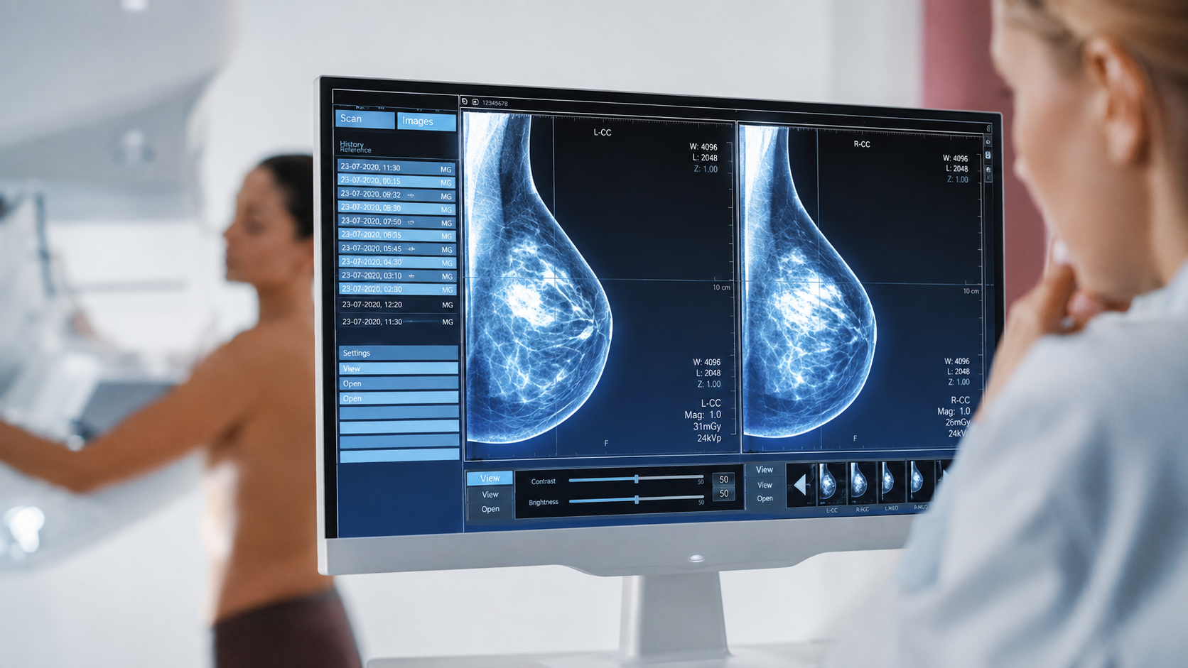
SonoMammography in OMR
Breast Ultrasound at AVR Scan Centre
If your doctor has recommended SonoMammography in OMR, you may want to understand what the examination is, why it may be recommended, whether it is painful, how to prepare, and how it differs from a mammogram.
SonoMammography, commonly referred to as breast ultrasound or breast sonography, uses high-frequency sound waves to create images of the internal structures of the breast.
It does not use ionizing radiation and is commonly used to evaluate breast lumps, pain, palpable abnormalities and findings seen on other breast examinations.
Breast ultrasound can help distinguish certain fluid-filled structures such as cysts from solid masses and can provide additional information about areas that may not be fully characterized by mammography alone.
AVR Scan Centre provides SonoMammography services for patients seeking breast imaging in and around OMR.
The right breast imaging examination depends on your age, symptoms, breast density, medical history, risk factors and the clinical question your doctor wants to answer.
What Is SonoMammography?
SonoMammography is an ultrasound examination of the breast that uses sound waves to create images of breast tissue and related structures.
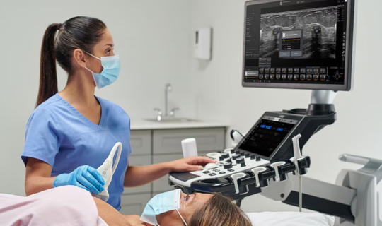
During the examination, a handheld ultrasound device called a transducer is placed over the breast with ultrasound gel between the device and the skin.
The transducer sends high-frequency sound waves into the breast and receives the returning echoes. A computer processes those signals into images that can be reviewed as part of the diagnostic assessment.
Breast ultrasound may provide information about:
- Breast tissue
- Breast lumps
- Cysts
- Solid masses
- Areas of tenderness or concern
- Selected lymph nodes
- Blood flow within a finding when Doppler is used
SonoMammography should be understood as breast ultrasound imaging, not as a replacement for every type of breast examination.
SonoMammography in OMR – Where Can You Get a Breast Ultrasound?
If you are searching for SonoMammography in OMR, AVR Scan Centre provides breast imaging services for patients looking for diagnostic care in and around the OMR region.
Patients from Sholinganallur, Padur, Navalur, Siruseri, Kelambakkam, Thiruporur and nearby OMR areas can enquire about the availability of the breast ultrasound examination recommended by their doctor.
Before your appointment, confirm:
- The exact breast examination required
- Whether ultrasound is being performed alone or with mammography
- Whether any preparation is required
- Appointment requirements
- Whether you should bring previous breast imaging reports
Because breast imaging recommendations differ according to the patient's age, symptoms and clinical history, it is better to confirm the exact examination before visiting.
Why Might a Doctor Recommend SonoMammography?
A doctor may recommend breast ultrasound when additional information about breast tissue or a particular breast finding is needed.
It may be used to evaluate:
- A breast lump
- Localized breast pain
- A palpable area of concern
- A finding seen on a mammogram
- A finding detected during a clinical breast examination
- Certain abnormalities in younger patients
- Selected breast findings that require further characterization
Breast ultrasound is often used as a diagnostic tool, especially when a specific area needs to be examined more closely.
The reason for the examination should be based on your clinical situation and your doctor's recommendation.
What Can a Breast Ultrasound Show?
Breast ultrasound can help characterize certain breast findings and may provide information that complements other breast imaging examinations.
Breast Lumps
Ultrasound can help examine a palpable breast lump and determine whether a finding has features of a fluid-filled cyst or a solid mass.
Breast Cysts
Some breast cysts can be visualized clearly on ultrasound.
Solid Masses
Ultrasound can characterize certain solid breast masses and may help determine whether additional investigation is appropriate.
Areas of Focal Pain
When breast pain is localized to a particular area, ultrasound may be used to investigate that region depending on the clinical assessment.
Findings Seen on Mammography
Ultrasound can provide additional information about a finding that is difficult to characterize on mammography alone.
Blood Flow
Doppler techniques may be used in selected situations to assess blood flow within a breast finding.
Is SonoMammography the Same as a Mammogram?
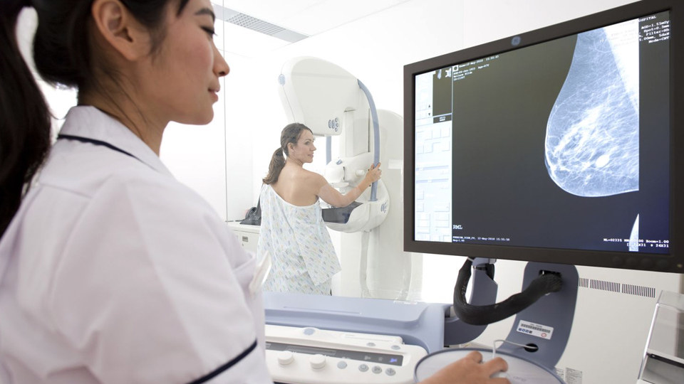
No. SonoMammography and mammography are different breast imaging examinations.
| SonoMammography / Breast Ultrasound | Mammography |
|---|---|
| Uses sound waves | Uses low-dose X-rays |
| Does not use ionizing radiation | Uses ionizing radiation |
| Provides ultrasound images of breast tissue | Produces X-ray images of the breast |
| Useful for evaluating certain lumps and abnormalities | Important for breast cancer screening and diagnosis |
| Can help distinguish some cystic and solid findings | Can show certain calcifications and other breast changes |
One test does not automatically replace the other. Depending on your age, symptoms, breast density and risk factors, your doctor may recommend ultrasound, mammography or both.
Breast ultrasound is often used as a complementary examination rather than a universal substitute for mammography.
Can Breast Ultrasound Replace a Mammogram?
No. Breast ultrasound should not generally be considered a complete replacement for mammography.
Ultrasound is useful for evaluating specific findings and can provide additional information in certain patients, including some women with dense breast tissue. However, mammography remains an important breast cancer screening tool.
The appropriate combination of breast imaging depends on:
- Age
- Symptoms
- Breast density
- Personal history
- Family history
- Risk level
- Previous imaging findings
- Doctor's recommendation
Is Breast Ultrasound Useful for Dense Breasts?
Breast ultrasound can provide additional information in some patients with dense breast tissue, but the decision to use it as supplemental imaging should be individualized.
Dense breast tissue can make some abnormalities harder to see on mammography. In selected patients, ultrasound may detect findings not visible on mammography.
However, ultrasound can also produce additional findings that turn out to be benign and may lead to further testing.
For this reason, supplemental breast imaging should be discussed with your doctor based on your individual risk and breast density rather than being treated as an automatic requirement for everyone.
Is SonoMammography Safe?
Breast ultrasound is non-invasive and does not use ionizing radiation.
Ultrasound uses sound waves to create images of the breast. Standard diagnostic breast ultrasound has no known harmful effects when appropriately performed.
It may be used in different patient groups when clinically appropriate, but the decision to perform any breast imaging should be based on the patient's medical needs.
Does SonoMammography Use Radiation?
No. SonoMammography does not use X-rays or ionizing radiation.
It uses:
High-frequency sound waves → Breast tissue images
This differs from:
- Mammography → Low-dose X-rays
- CT → X-rays
- MRI → Magnetic fields and radio waves
The absence of radiation is one of the key characteristics of breast ultrasound.
Is a Breast Ultrasound Painful?
Breast ultrasound is generally non-invasive and is usually not painful.
During the examination, a transducer is moved over the breast with gel applied to the skin.
You may feel some pressure as the transducer is positioned over the area being examined.
If your breast is already tender or painful, the pressure may cause temporary discomfort. Inform the healthcare professional if the examination causes significant discomfort.
How Should You Prepare for SonoMammography?
Breast ultrasound usually requires little to no special preparation.
Wear Comfortable Clothing
Wear clothing that can be easily removed from the upper body when required for the examination.
Avoid Excessive Jewellery
You may be asked to remove jewellery or other items that interfere with the examination.
Bring Previous Breast Reports
Previous mammograms, ultrasounds, breast MRI reports or other relevant records can provide useful clinical context when available.
Tell the Healthcare Team About Your Symptoms
- A new lump
- Breast pain
- Nipple changes
- Skin changes
- Previous breast surgery
- Previous breast imaging findings
- Family or personal history of breast disease
Your doctor can determine whether additional imaging is required.
What Happens During a SonoMammography Scan?
A breast ultrasound is generally a straightforward examination.
1. Preparation
You will usually be asked to remove clothing from the upper body and wear a gown.
2. Positioning
You will lie on an examination table, generally on your back, and may be asked to raise an arm depending on the area being examined.
3. Gel Application
Ultrasound gel is applied to the skin of the breast.
4. Scanning
The transducer is moved across the breast to obtain images of the required areas.
5. Additional Views
The healthcare professional may examine the area from different directions or use Doppler imaging when appropriate.
6. Completion
Once the required images have been obtained, the gel is removed and the examination is completed.
Can Breast Ultrasound Detect Breast Cancer?
Breast ultrasound can help evaluate breast abnormalities and may identify some cancers, but ultrasound alone cannot determine that every breast lump is cancerous or rule out every breast cancer.
Ultrasound can help characterize certain breast findings and may identify abnormalities that are not clearly visible on mammography.
If an ultrasound finding appears suspicious, your doctor may recommend further evaluation, which could include additional breast imaging or a biopsy depending on the circumstances.
A breast ultrasound report should be interpreted by an appropriately qualified healthcare professional in the context of your clinical history.
Can Breast Ultrasound Tell If a Lump Is Benign?
Ultrasound can help characterize some breast lumps, but imaging alone does not always provide a definitive diagnosis.
For example, ultrasound can help distinguish a simple fluid-filled cyst from a solid mass in many cases.
Some findings may require:
- Additional imaging
- Follow-up ultrasound
- Mammography
- Breast MRI
- Image-guided biopsy
The next step depends on the appearance of the finding and the clinical assessment.
What Happens If the Ultrasound Finds a Breast Abnormality?
An abnormal ultrasound finding does not automatically mean breast cancer.
Many breast findings are benign.
Depending on the imaging appearance, your doctor may recommend:
- Routine follow-up
- Repeat ultrasound
- Mammography
- Additional breast imaging
- Biopsy
- Specialist consultation
Additional testing is sometimes necessary because ultrasound is one part of a broader breast diagnostic process.
What Is Doppler Breast Ultrasound?
Doppler ultrasound is a specialized ultrasound technique that can evaluate blood flow within blood vessels or, in selected cases, within a breast finding.
It may provide additional information about vascularity when clinically appropriate.
Doppler is not required for every breast ultrasound.
Breast Ultrasound vs Breast MRI – What Is the Difference?
| Breast Ultrasound | Breast MRI |
|---|---|
| Uses sound waves | Uses a strong magnetic field and radio waves |
| Does not use ionizing radiation | Does not use ionizing radiation |
| Useful for evaluating certain lumps and abnormalities | Provides detailed breast imaging for selected indications |
| Can distinguish certain cystic and solid findings | Useful for selected high-risk screening and diagnostic situations |
| Usually does not require contrast | Many breast MRI examinations use contrast |
Your doctor determines which examination is appropriate based on your symptoms, risk factors and the clinical question.
Can SonoMammography Be Done During Pregnancy?
Breast ultrasound can be used during pregnancy when breast imaging is clinically required.
Because ultrasound does not use ionizing radiation, it can be a useful imaging method when a breast concern needs to be evaluated during pregnancy.
However, breast symptoms during pregnancy should still be assessed by a healthcare professional, who can determine whether ultrasound alone is sufficient or whether other imaging is needed.
What Happens After a SonoMammography Scan?
After the examination, the breast ultrasound images are reviewed as part of the diagnostic process.
The findings may be considered along with:
- Your symptoms
- Physical examination
- Age
- Breast density
- Medical history
- Previous mammograms
- Previous ultrasound reports
- Family or personal history of breast disease
Your doctor can explain what the findings mean and whether further evaluation is necessary.
Do not try to diagnose a breast condition based on ultrasound images alone.
Why Choose AVR Scan Centre for SonoMammography in OMR?
When looking for a SonoMammography Centre in OMR, patients may want to consider the availability of breast ultrasound, location, appointment requirements and access to related breast imaging services.
AVR Scan Centre provides SonoMammography services for patients seeking breast imaging in and around OMR.
1. SonoMammography Service
AVR Scan Centre offers SonoMammography as part of its diagnostic imaging services.
2. OMR-Side Accessibility
The centre's OMR location makes it relevant for patients looking for diagnostic services around the OMR region.
3. Patient Preparation Guidance
Patients can contact the centre before their appointment to confirm the required breast examination and any instructions for the visit.
4. Related Breast Imaging Services
AVR Scan Centre also provides other breast imaging services, including:
- Digital Mammogram
- SonoMammography
Patients can discuss with their doctor which breast imaging examination is appropriate for their individual situation.
What Should You Check Before Booking SonoMammography in OMR?
Before scheduling a breast ultrasound, confirm:
- Why your doctor has recommended the examination
- Whether the examination is for a specific lump or symptom
- Whether you need ultrasound alone or ultrasound with mammography
- Whether previous breast imaging is available
- Whether any preparation is required
- Whether an appointment is needed
Before You Visit AVR Scan Centre
Bring your doctor's prescription or referral and any previous breast imaging reports that may help with comparison.
If you have noticed a new breast lump, persistent focal breast pain, nipple discharge, skin changes or another breast concern, discuss it with a qualified healthcare professional rather than relying only on an imaging test.
Frequently Asked Questions About SonoMammography in OMR
SonoMammography is an ultrasound examination of the breast that uses high-frequency sound waves to create images of breast tissue.
AVR Scan Centre provides SonoMammography services for patients looking for breast imaging in and around the OMR region. Contact the centre to confirm availability and appointment details.
No. Breast ultrasound uses sound waves and does not use ionizing radiation.
Breast ultrasound is generally non-invasive and usually not painful, although some pressure may be felt while the transducer is moved over the breast.
Usually, no special preparation is needed for breast ultrasound.
Yes. Breast ultrasound can help evaluate a breast lump and may help distinguish some fluid-filled cysts from solid masses.
Breast ultrasound can identify and characterize some suspicious breast findings, including some cancers, but it cannot detect every breast cancer and does not replace appropriate breast screening.
No. Breast ultrasound is often used as a complementary examination rather than a universal replacement for mammography.
It can provide additional information in selected patients with dense breast tissue, although the decision to use supplemental ultrasound should be individualized because additional findings and false positives can occur.
SonoMammography uses ultrasound sound waves, while mammography uses low-dose X-rays. They provide different types of breast imaging information.
Breast ultrasound uses sound waves, while breast MRI uses a magnetic field and radio waves. They are used for different clinical indications.
Yes. Breast ultrasound can be used during pregnancy when clinically indicated. Your healthcare professional will determine the appropriate imaging approach.
Ultrasound can characterize some benign findings, such as certain simple cysts, but not every breast lesion can be definitively classified by ultrasound alone.
Your doctor may recommend follow-up imaging, mammography, another imaging examination or biopsy depending on the finding and clinical situation.
Bring your doctor's prescription or referral and previous breast imaging reports when available.
The duration depends on the number of areas being examined and whether additional views or Doppler assessment are needed. Ask the centre about the expected duration of your specific examination.
Persistent or localized breast pain should be discussed with a healthcare professional. Depending on your age, symptoms and clinical findings, your doctor may recommend ultrasound, mammography or another examination.
Yes. A new or persistent breast lump should be assessed by a qualified healthcare professional. Depending on your age and clinical findings, breast imaging may be recommended.
Get Your SonoMammography at AVR Scan Centre, Sholinganallur
Breast ultrasound can provide valuable information when a doctor needs to investigate a breast lump, pain, imaging finding or other breast concern.
Understanding why the examination has been recommended and whether you need ultrasound alone or alongside another breast imaging test can help you prepare for your appointment.
AVR Scan Centre provides SonoMammography services for patients looking for breast imaging in and around OMR.
Before visiting, contact the centre to confirm:
- Your required breast imaging examination
- Appointment availability
- Preparation instructions
- Whether previous breast imaging should be brought
- Any specific instructions for your appointment
Need a SonoMammography in OMR?
Contact AVR Scan Centre – Sholinganallur to enquire about the breast ultrasound recommended by your doctor and confirm appointment availability.
Contact AVR Scan Centre – Sholinganallur to enquire about the breast ultrasound recommended by your doctor and confirm appointment availability.
AVR Scan Centre – Sholinganallur
First Floor, No. 278/1 & 278/2, Commercial Building,
Ramanyam Pushkar 2, Kalaignar Karunanidhi Salai,
Sholinganallur – 600119.
