

Digital Mammogram in OMR
Digital Breast Imaging at AVR Scan Centre
If you are looking for a Digital Mammogram in OMR, you may have questions about what a mammogram checks, who may need one, how the examination is performed, whether it is painful, how to prepare and how mammography differs from breast ultrasound.
A digital mammogram is a breast imaging examination that uses low-dose X-rays to produce digital images of the breast. Mammography is an important tool for breast cancer screening because it can detect some breast abnormalities, including certain cancers, before they can be felt as a lump.
During a mammogram, the breast is briefly compressed between plates so that the breast tissue can be spread out and imaged clearly.
AVR Scan Centre provides Digital Mammogram services for patients looking for breast imaging in and around the OMR region.
The right mammography schedule and whether screening or diagnostic imaging is appropriate depend on factors such as age, symptoms, personal history, family history, breast density and individual breast-cancer risk.
What Is a Digital Mammogram?
A digital mammogram is a low-dose X-ray examination that produces digital images of the breast for screening or diagnostic evaluation.
Instead of producing an image on traditional film, digital mammography captures the X-ray information electronically and displays the images digitally.
A mammogram may help detect:
- Certain breast masses
- Some small breast abnormalities
- Microcalcifications
- Other changes that may warrant further assessment
Mammography is used for both screening and diagnostic evaluation, but these are not the same.
Screening Mammogram
A screening mammogram is performed in people who do not have a specific breast symptom or known abnormality and is used to look for signs of breast cancer at an earlier stage.
Diagnostic Mammogram
A diagnostic mammogram may be recommended when there is a breast symptom, a new lump, a breast change, or an abnormality found on previous breast imaging.
The exact examination should be determined by your doctor or breast-imaging team.
Digital Mammogram in OMR – Where Can You Get a Mammogram?
If you are searching for a Digital Mammogram in OMR, AVR Scan Centre provides Digital Mammogram services for patients seeking breast imaging in and around the OMR region.
The Sholinganallur centre serves patients from OMR and nearby areas. Patients travelling from Padur, Navalur, Siruseri, Kelambakkam, Thiruporur and surrounding localities can contact AVR Scan Centre before visiting to confirm the availability of the required mammography examination.
Before your appointment, confirm:
- Whether you need a screening or diagnostic mammogram
- Appointment requirements
- Preparation instructions
- Whether you should bring previous breast imaging
- Whether your doctor has requested additional breast imaging
AVR Scan Centre – Sholinganallur
First Floor, No. 278/1 & 278/2, Commercial Building, Ramanyam Pushkar 2, Kalaignar Karunanidhi Salai, Sholinganallur – 600119.
Why Might a Doctor Recommend a Digital Mammogram?
A doctor may recommend a mammogram for breast cancer screening or to investigate a breast symptom or imaging finding.
A mammogram may be considered when:
- Routine breast screening is due according to your individualized screening plan
- A breast lump has been noticed
- There is a change in breast shape or size
- There are skin changes
- There is nipple discharge or another nipple change
- An earlier breast examination showed a finding requiring further assessment
- Previous breast imaging showed an area that needs additional evaluation
A mammogram can detect some breast cancers before they are large enough to be felt.
The decision about when to start and how often to have screening should take into account your age, risk factors and local clinical guidance rather than applying the same schedule to everyone.
What Can a Digital Mammogram Show?
A digital mammogram can show certain breast abnormalities that may not be detectable by physical examination alone.
Depending on the examination, mammography can help evaluate:
Breast Masses
A mammogram may show a mass or other area of concern within the breast tissue.
Calcifications
Mammography is particularly useful for detecting small calcifications, including patterns that may require further evaluation.
Changes in Breast Tissue
Mammograms can show changes in breast tissue that may be difficult to detect by physical examination.
Differences Compared With Previous Images
When previous mammograms are available, comparison may help the interpreting specialist identify whether a finding is new, unchanged or has changed over time.
A mammogram does not automatically diagnose breast cancer. An abnormal finding may require additional imaging, follow-up or biopsy depending on the result and clinical context.
What Is the Difference Between a Screening and Diagnostic Mammogram?
A screening mammogram is used to look for breast cancer in people without a specific breast complaint, while a diagnostic mammogram is used to investigate a symptom or an abnormal finding.
| Screening Mammogram | Diagnostic Mammogram |
|---|---|
| Usually performed when there is no specific breast symptom | Performed when a symptom or abnormality requires evaluation |
| Focuses on early detection | Focuses on investigating a specific concern |
| Usually follows a planned screening schedule | May be recommended outside routine screening |
| Additional views may be needed if something is seen | Additional targeted imaging is often part of the evaluation |
The appropriate type of mammogram depends on your individual situation.
How Does a Digital Mammogram Work?
Digital mammography uses low-dose X-rays to create digital images of breast tissue.
During the examination, the breast is positioned on a mammography unit and briefly compressed between two surfaces. Compression spreads the breast tissue and reduces overlapping structures, helping produce clearer images.
The X-ray information is captured digitally and displayed for interpretation.
Different views of each breast are commonly obtained to provide a more complete assessment.
What Happens During a Digital Mammogram?
A mammogram is a relatively quick examination, but the positioning process is important for obtaining useful images.
1. Changing for the Examination
You will generally need to remove clothing from the upper body and wear the appropriate gown or follow the facility's instructions.
2. Breast Positioning
A mammography technologist positions the breast carefully on the imaging platform.
3. Breast Compression
The breast is compressed for a few seconds while an image is acquired. Compression helps spread the tissue and reduce overlap.
4. Multiple Images
Several views may be taken as required.
5. Completion
Once the necessary images have been obtained, the examination is completed. The images are then reviewed as part of the breast-imaging process.
Is a Digital Mammogram Painful?
Mammography is not usually painful, but breast compression can feel uncomfortable for some people.
The compression is brief and is important for obtaining good-quality images while limiting tissue overlap.
If you experience significant pain during the examination, tell the technologist. Communication can help with positioning and comfort.
Breast tenderness may also vary at different times of the menstrual cycle.
How Should You Prepare for a Digital Mammogram?
Most people do not need to fast before a mammogram.
Avoid Deodorant or Certain Products on the Breast Area
Do not use deodorant, talcum powder, lotion or similar products around the underarms or breasts unless the imaging centre advises otherwise, because some products can contain substances that may appear on mammography images.
Wear Comfortable Clothing
Wear a two-piece outfit if that makes it easier to change for the examination.
Tell the Mammography Team About Breast Implants
Inform the team if you have breast implants so appropriate positioning and imaging techniques can be used.
Tell Them If You Are Pregnant or May Be Pregnant
Because mammography uses X-rays, inform your healthcare professional and the imaging team if you are pregnant or think you may be pregnant.
Bring Previous Mammograms
Previous mammograms and breast imaging reports can be valuable for comparison, especially when you are changing centres.
Does a Digital Mammogram Use Radiation?
Yes. Digital mammography uses a small dose of X-ray radiation to produce breast images.
The radiation dose is kept low while obtaining diagnostically useful images.
Mammography is used because its potential benefit in detecting breast cancer early needs to be considered alongside the small radiation exposure associated with the examination.
If you are pregnant or may be pregnant, tell the healthcare team before the examination.
Is a Digital Mammogram Safe?
Mammography is an established breast-imaging examination that uses a low dose of X-ray radiation.
Quality mammography depends on appropriate equipment, image quality, trained personnel and proper examination procedures.
Like any X-ray examination, mammography involves radiation exposure, so screening should follow an appropriate clinical plan.
For people at higher risk of breast cancer, the appropriate screening approach may differ from routine screening. Discuss your personal risk with your healthcare professional.
What Is Breast Density and Why Does It Matter?
Breast density describes the relative amount of fibrous and glandular tissue compared with fatty tissue in the breasts as seen on a mammogram.
Dense breast tissue can make some abnormalities harder to see on mammography and can also be associated with an increased risk of breast cancer. The significance of breast density varies among individuals.
Whether additional imaging such as ultrasound or MRI is appropriate depends on your personal risk, breast density, previous imaging and local clinical guidance.
The USPSTF has stated that evidence is currently insufficient to determine the overall balance of benefits and harms of supplemental ultrasound or MRI screening solely on the basis of dense breasts.
That means additional imaging should not automatically be assumed to be necessary for everyone with dense breasts.
Digital Mammogram vs SonoMammography – What Is the Difference?
Digital mammography and breast ultrasound use different imaging technologies and can provide complementary information.
| Digital Mammogram | SonoMammography / Breast Ultrasound |
|---|---|
| Uses low-dose X-rays | Uses sound waves |
| Particularly important for breast cancer screening | Commonly used to evaluate specific breast findings |
| Can show certain calcifications | Can help characterize some cystic and solid findings |
| Produces mammographic X-ray images | Produces ultrasound images |
| Breast compression is required | No breast compression in the same way as mammography |
Neither test universally replaces the other.
Breast ultrasound may be recommended in addition to mammography when a specific finding needs further evaluation or in selected clinical situations.
Can a Digital Mammogram Detect Breast Cancer Early?
Yes. Mammography can detect some breast cancers at an early stage, including abnormalities that may be too small to feel.
Early detection may provide more treatment options, but a mammogram does not find every breast cancer.
A normal mammogram also does not mean that future breast changes should be ignored.
Contact a healthcare professional if you notice a new breast lump or another persistent breast change even if your most recent mammogram was normal.
Does a Normal Digital Mammogram Rule Out Breast Cancer?
No. A normal mammogram cannot rule out every breast cancer or every breast abnormality.
Mammography can miss some abnormalities, particularly in certain clinical situations.
If you notice:
- A new breast lump
- Skin changes
- Nipple changes
- Persistent focal breast symptoms
- Nipple discharge
- Any other concerning breast change
you should discuss it with a healthcare professional even if your previous mammogram was normal.

What Happens If a Mammogram Shows an Abnormality?
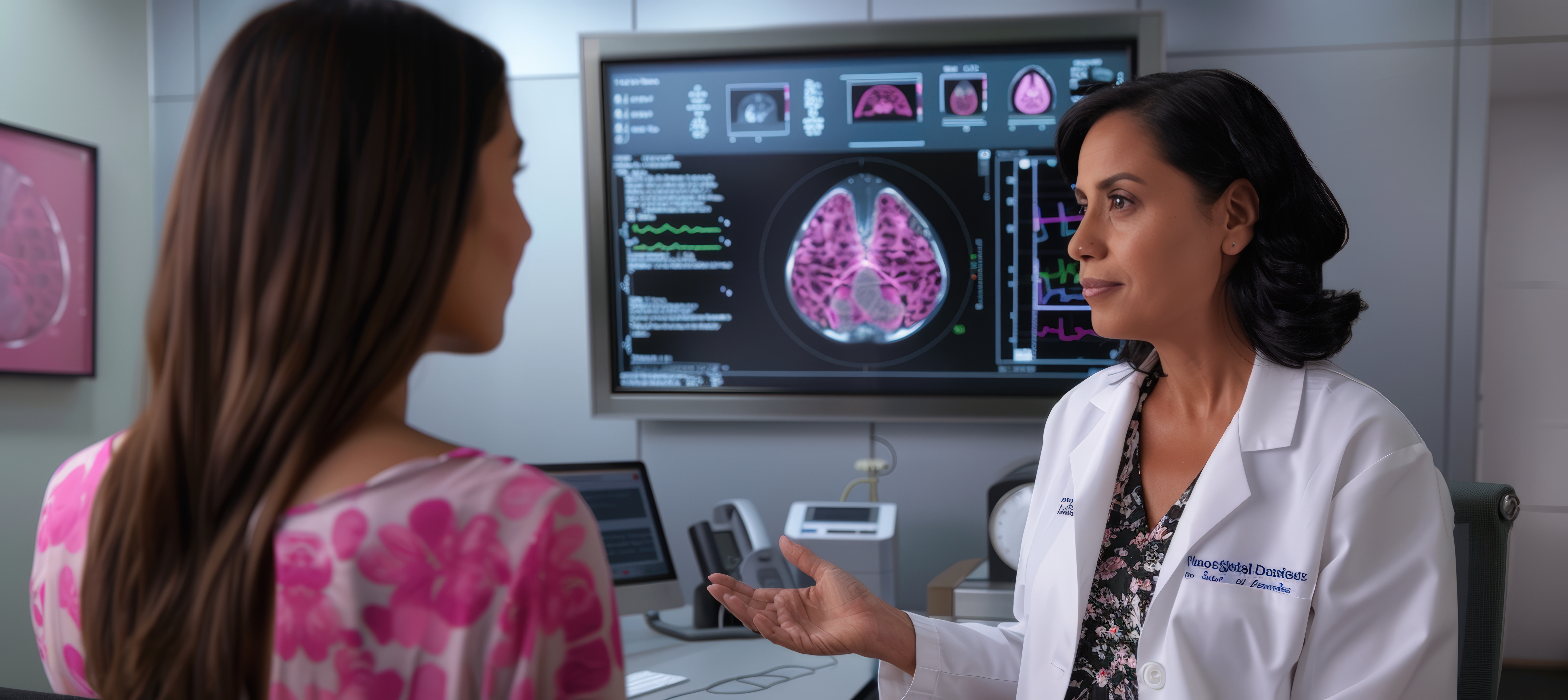
An abnormal mammogram does not automatically mean that you have breast cancer.
Many mammography findings require additional evaluation before a diagnosis can be made.
Depending on the finding, your doctor or breast-imaging team may recommend:
- Additional mammographic views
- Breast ultrasound
- Follow-up imaging
- Breast MRI in selected situations
- Image-guided biopsy
The next step depends on the appearance of the finding and your clinical history.
Do not interpret a mammogram result based on one word or one image alone.
Can I Have a Digital Mammogram With Breast Implants?
Women with breast implants can generally undergo mammography, but the imaging team needs to know about the implants before the examination.
Additional or modified views may be required to visualize as much breast tissue as possible.
Tell the mammography team:
- That you have implants
- Whether the implants are saline or silicone, if known
- About any previous breast surgery
- About any concerns related to the implants
The exact imaging approach will be determined by the trained mammography team.
What Is the Difference Between Digital Mammography and 3D Mammography?
Digital mammography generally refers to conventional digital mammography, while 3D mammography usually refers to digital breast tomosynthesis (DBT).
Digital mammography creates conventional 2D breast images. 3D tomosynthesis acquires multiple X-ray projections from different angles and reconstructs them into thin image sections.
Both digital mammography and digital breast tomosynthesis are recognized mammographic screening modalities.
Do not assume that “3D” is automatically better for every patient. The appropriate breast imaging method depends on the clinical situation, available technology and screening recommendation.
When Should I Get a Digital Mammogram?
The appropriate timing of breast screening depends on your age, personal risk, family history, previous breast findings and the screening recommendations followed by your healthcare provider.
There is no single screening interval that is appropriate for every person.
As one example, the 2024 USPSTF recommendation advises screening mammography every two years for women aged 40 to 74 at average risk. However, recommendations differ among organizations and countries, and people at higher risk may need a different approach.
For patients in India, the most appropriate screening schedule should be discussed with a qualified healthcare professional who can consider your individual risk and local clinical practice.
What Happens After a Digital Mammogram?
After your mammography images are obtained, they are reviewed as part of the breast-imaging process.
The interpretation may take into account:
- Your age
- Symptoms
- Breast density
- Previous mammograms
- Previous breast ultrasound
- Personal history
- Family history
- Other relevant clinical information
If an abnormality is identified, additional imaging or follow-up may be recommended.
A mammogram report should therefore be interpreted in its complete clinical context rather than through self-reading of the images.
Why Choose AVR Scan Centre for a Digital Mammogram in OMR?
When searching for a Digital Mammogram in OMR, patients may want to consider the availability of mammography, location, preparation instructions, appointment process and access to related breast-imaging services.
AVR Scan Centre provides Digital Mammogram services for patients seeking breast imaging around the OMR region.
1. Digital Mammography Service
AVR Scan Centre provides Digital Mammogram services as part of its diagnostic imaging offering.
2. OMR-Side Accessibility
The Sholinganallur centre serves patients looking for diagnostic services in and around the OMR region.
3. Patient Preparation Guidance
Patients can contact the centre before their appointment to confirm the examination process and any preparation instructions.
4. Related Breast Imaging
AVR Scan Centre also provides SonoMammography, allowing patients to discuss the appropriate breast imaging examination based on their doctor's recommendation.
What Should You Check Before Booking a Digital Mammogram in OMR?
Before scheduling your mammogram, confirm:
- Whether the examination is for screening or a specific breast concern
- Whether you have previous mammograms available for comparison
- Whether you have breast implants
- Whether you are pregnant or may be pregnant
- Whether the centre has specific preparation instructions
- Whether an appointment is required
Before You Visit AVR Scan Centre
Bring your doctor's prescription or referral where applicable and previous breast imaging reports or mammograms when available.
If you have a new breast lump, persistent breast change, nipple discharge, skin change or another breast concern, discuss it with a healthcare professional rather than waiting for routine screening.
Frequently Asked Questions About Digital Mammogram in OMR
A digital mammogram is a low-dose X-ray examination that creates digital images of the breast for screening or diagnostic evaluation.
AVR Scan Centre provides Digital Mammogram services for patients looking for breast imaging in and around the OMR region. Contact the centre to confirm availability and appointment details.
Yes. Mammography uses a small dose of X-ray radiation to produce breast images.
A mammogram may cause temporary pressure or discomfort because the breast needs to be compressed for image acquisition. The compression usually lasts only a few seconds per image.
Generally, fasting is not required for a routine mammogram.
It is usually recommended to avoid deodorant, talcum powder, lotion or similar products on the breast and underarm area before mammography because some products can create appearances on the images that may interfere with interpretation.
Yes, mammography can generally be performed in people with breast implants, but tell the mammography team about the implants before the examination because additional or modified views may be required.
Tell your doctor and mammography team if you are pregnant or may be pregnant. Because mammography uses X-rays, the healthcare team will determine whether imaging is appropriate and how to proceed.
No. Conventional digital mammography produces 2D images, while 3D mammography generally refers to digital breast tomosynthesis, which reconstructs thin image sections from multiple X-ray projections.
Mammography can detect some breast cancers and other suspicious breast changes, including findings that may be too small to feel. However, mammography does not detect every breast cancer.
No. A normal mammogram does not rule out every breast cancer. New or persistent breast changes should still be assessed by a healthcare professional.
An abnormal result does not automatically mean cancer. Additional mammographic views, ultrasound, follow-up imaging or biopsy may be recommended depending on the finding.
Neither is universally better. Mammography and breast ultrasound use different technologies and have different roles. Ultrasound is often used as a complementary diagnostic examination.
Yes, mammography remains an important breast-imaging tool in people with dense breasts, but dense tissue can make some abnormalities harder to see. Whether additional imaging is appropriate depends on individual risk and clinical circumstances.
There is no one schedule suitable for everyone. Recommendations vary by country, age, personal risk and the organization providing the guideline. Discuss the appropriate screening schedule with your healthcare professional.
The appropriate interval depends on age, breast-cancer risk, previous findings and the screening guideline being followed. Your healthcare professional can advise you on the interval appropriate for you.
Bring your doctor's referral if applicable and previous mammograms or breast imaging reports if available. Tell the mammography team about breast implants and relevant breast history.
The actual imaging process is usually brief, although total appointment time varies with registration, positioning, image acquisition and whether additional views are required.
Get Your Digital Mammogram at AVR Scan Centre, Sholinganallur
A digital mammogram is an important breast-imaging examination that can help detect certain breast abnormalities and support early detection of breast cancer.
Understanding whether you need screening or diagnostic mammography, how to prepare and what to expect from breast compression can make your appointment easier.
AVR Scan Centre provides Digital Mammogram services for patients looking for breast imaging in and around OMR.
Before visiting, contact the centre to confirm:
- Your required mammography examination
- Appointment availability
- Preparation instructions
- Whether you should bring previous breast imaging
- Any specific instructions related to your appointment
Contact AVR Scan Centre – Sholinganallur to enquire about the mammogram recommended by your doctor and confirm appointment availability.
AVR Scan Centre – Sholinganallur
First Floor, No. 278/1 & 278/2, Commercial Building,
Ramanyam Pushkar 2, Kalaignar Karunanidhi Salai,
Sholinganallur – 600119.
