
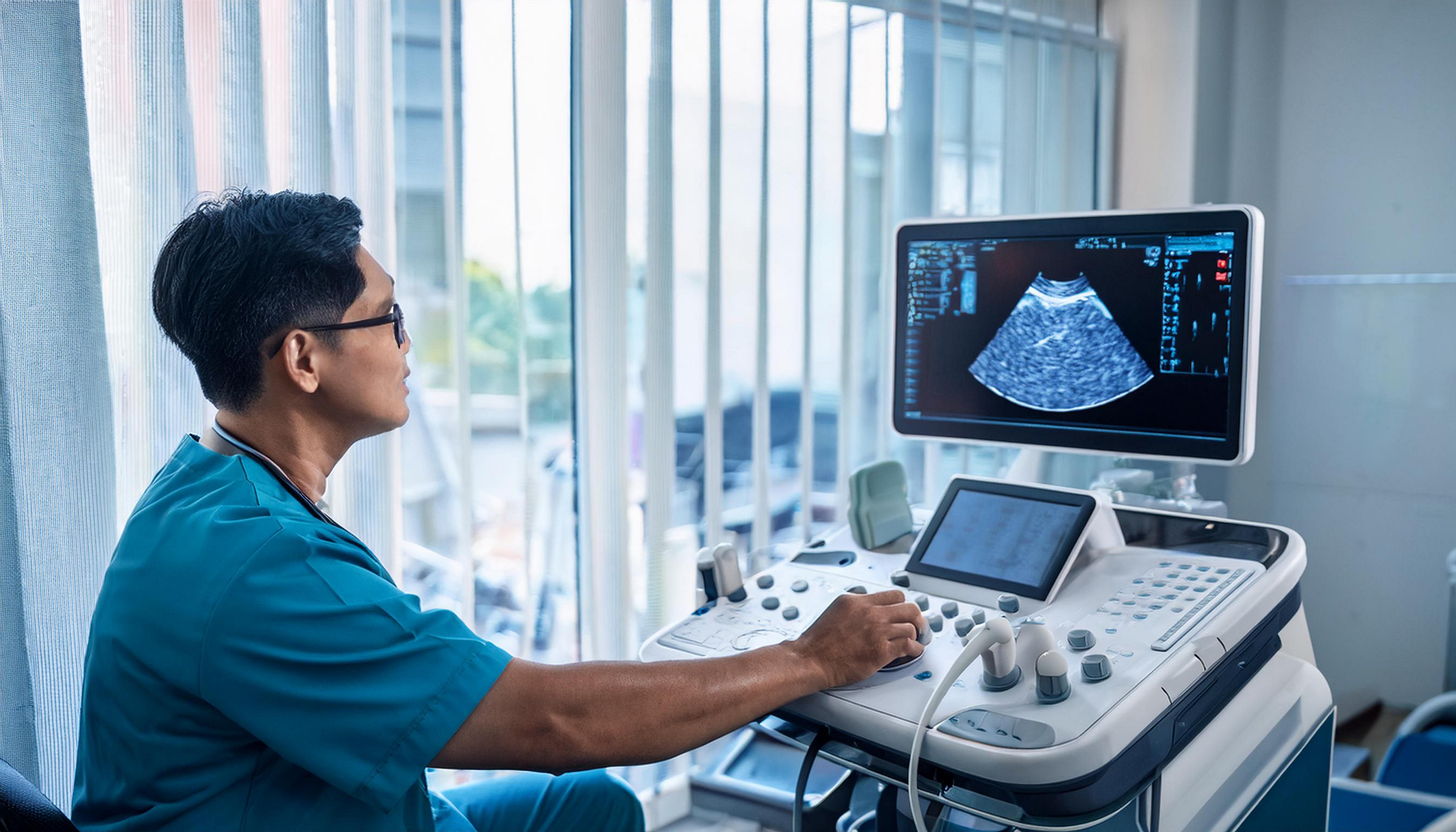
Echocardiogram in OMR
Echo Test at AVR Scan Centre
If your doctor has recommended an Echocardiogram in OMR, you may have questions about what an Echo test examines, why it has been recommended, whether you need to prepare, whether it is painful, and what the results can tell your doctor.
An echocardiogram, also called an Echo or heart ultrasound, is a non-invasive test that uses high-frequency sound waves to create moving images of the heart. It can provide information about the heart's structure, pumping function, valves, chambers and blood flow.
AVR Scan Centre provides Echocardiogram services for patients looking for cardiac diagnostic testing in and around the OMR region.
The specific type of echocardiogram you need depends on your symptoms, medical history and the clinical question your doctor wants to evaluate.
What Is an Echocardiogram?
An echocardiogram is a heart ultrasound examination that uses sound waves to create moving images of the heart.
Unlike CT scans and X-rays, a standard echocardiogram does not use ionizing radiation. The examination can show how the heart moves and pumps blood and can provide information about the heart's chambers, walls and valves.
An echocardiogram may help healthcare professionals assess:
- Size and shape of the heart
- Heart muscle movement
- Pumping function
- Heart valves
- Blood flow through the heart
- Certain structural abnormalities
- Fluid around the heart
- Selected congenital heart problems
The findings need to be interpreted in the context of your symptoms, medical history and other clinical information.
Echocardiogram in OMR – Where Can You Get an Echo Test?
If you are searching for an Echocardiogram in OMR, AVR Scan Centre provides Echo Test services for patients looking for cardiac diagnostic imaging around the OMR region.
The Sholinganallur centre serves patients from OMR and nearby localities.
Patients travelling from Padur, Navalur, Siruseri, Kelambakkam, Thiruporur and surrounding areas can contact AVR Scan Centre before visiting to confirm the availability of the echocardiogram recommended by their doctor.
AVR Scan Centre – Sholinganallur:
First Floor, No. 278/1 & 278/2, Commercial Building,
Ramanyam Pushkar 2, Kalaignar Karunanidhi Salai,
Sholinganallur – 600119.
Before your appointment, confirm:
- The type of Echo test required
- Appointment availability
- Whether any specific preparation is needed
- Whether you should bring previous cardiac reports
- Any instructions related to medications
Why Might a Doctor Recommend an Echocardiogram?
A doctor may recommend an echocardiogram when information about the heart's structure, movement, pumping function or blood flow is needed.
An Echo may be considered when evaluating symptoms or clinical findings such as:
- Chest discomfort
- Shortness of breath
- Heart murmur
- Swelling related to suspected heart problems
- Palpitations in selected clinical situations
- Fainting or unexplained symptoms
- Suspected valve disease
- Suspected heart failure
- Known or suspected structural heart disease
An echocardiogram may also be used to monitor a known heart condition or assess the heart before or after certain treatments when clinically appropriate. The reason for the test depends on your individual clinical situation.
What Can an Echocardiogram Show?
An echocardiogram can provide information about the heart's structure and how well it is functioning.
Heart Size and Chambers
An echocardiogram can provide information about the size and shape of the heart and its chambers.
Heart Muscle Movement
The examination can show how different parts of the heart muscle move during each heartbeat. Areas of reduced movement may require further clinical assessment.
Pumping Function
An echocardiogram can help assess how effectively the heart pumps blood.
One measurement that may be reported is the ejection fraction, which describes the proportion of blood pumped out of a heart chamber with each contraction.
Heart Valves
Echo can help assess whether heart valves open and close normally and whether there is narrowing or leakage. Valve narrowing is called stenosis, while backward leakage is called regurgitation.
Blood Flow
Doppler techniques can provide information about the direction and speed of blood flow through the heart and valves.
Fluid Around the Heart
An echocardiogram can help identify fluid around the heart, known as a pericardial effusion, when present.
What Is a 2D Echocardiogram?
A 2D echocardiogram uses ultrasound to produce two-dimensional moving images of the heart.
It can allow healthcare professionals to assess:
- Heart chambers
- Heart walls
- Heart valves
- Heart muscle movement
- Overall cardiac structure
The standard transthoracic echocardiogram (TTE) is performed by placing an ultrasound transducer on the chest. The exact measurements and views obtained depend on the patient's condition and the Echo protocol.
What Is Doppler Echocardiography?
Doppler echocardiography is an ultrasound technique that evaluates blood flow through the heart and major blood vessels.
Doppler information can help healthcare professionals assess:
- Direction of blood flow
- Speed of blood flow
- Valve narrowing
- Valve leakage
- Certain abnormal flow patterns
- Selected pressure-related measurements
Doppler can complement structural images and provide functional information about how blood moves through the heart.
What Are the Different Types of Echocardiograms?
The type of echocardiogram depends on the clinical question and the information your doctor needs.
Transthoracic Echocardiogram
A transthoracic echocardiogram (TTE) is the standard form of Echo. The ultrasound transducer is placed on the chest to create images of the heart.
Doppler Echocardiogram
Doppler techniques provide information about blood flow through the heart and valves.
Stress Echocardiogram
A stress echocardiogram evaluates the heart while it is under physical or pharmacological stress and may be used for selected clinical questions.
Transesophageal Echocardiogram
A transesophageal echocardiogram (TEE/TOE) uses a specialized probe passed into the esophagus to obtain detailed views of the heart from close to it. It is a different procedure from a routine transthoracic Echo and requires specific preparation and medical supervision.
3D Echocardiogram
Three-dimensional echocardiography can provide additional structural views in selected situations.
Not every patient needs every type of echocardiogram. The appropriate examination is selected according to the clinical requirement.
How Does an Echocardiogram Work?
An echocardiogram uses sound waves to create moving images of the heart.
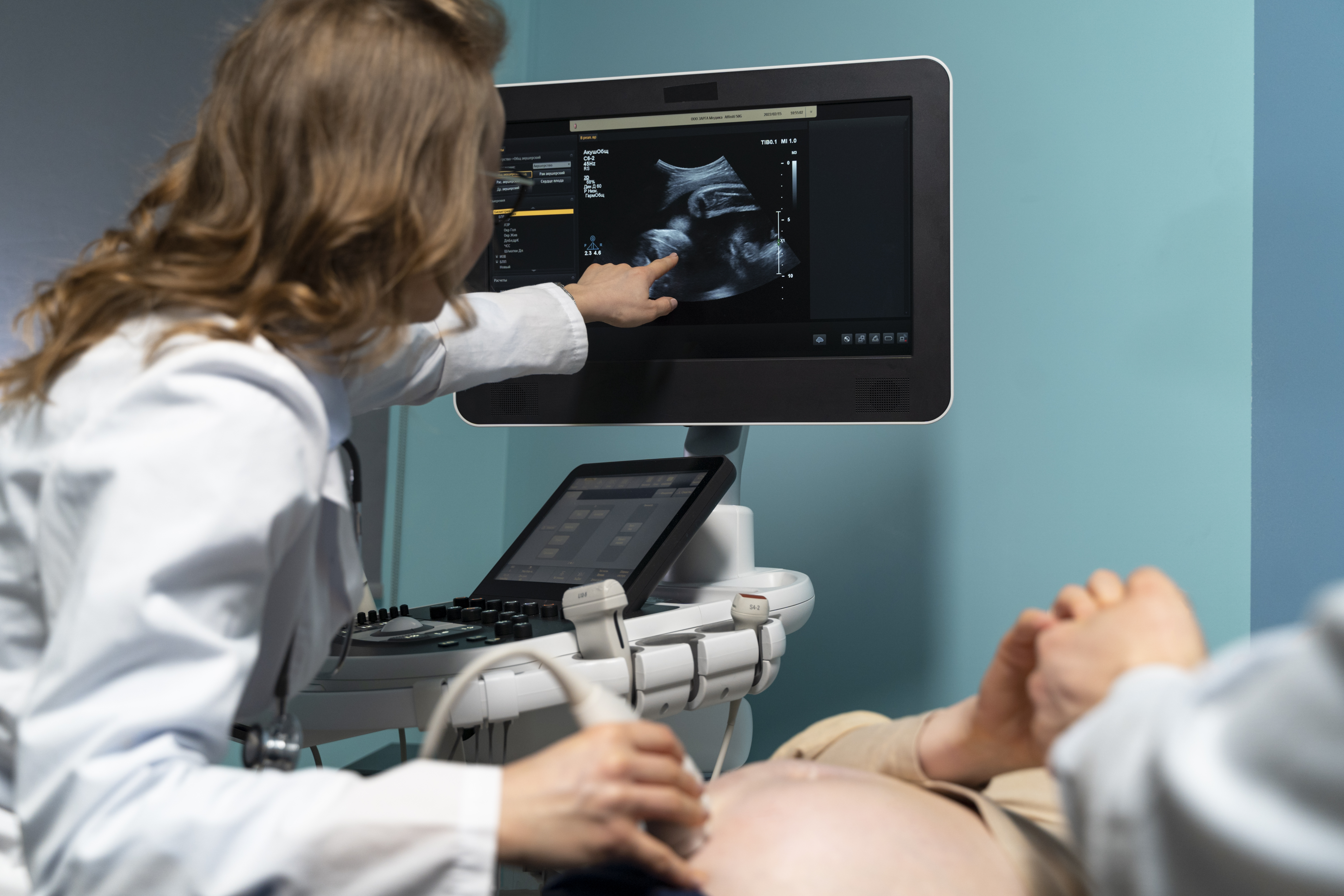
During a standard Echo, a handheld ultrasound transducer is placed against the chest with gel between the transducer and skin.
The transducer sends sound waves into the body and detects returning echoes. A computer converts these signals into images that can be viewed on a monitor.
The examination can provide real-time information about the movement of the heart and, with Doppler techniques, blood flow.
What Happens During an Echocardiogram?
A standard transthoracic Echo is generally a non-invasive examination.
1. Positioning
You will usually lie on an examination bed, often turned slightly onto your left side.
2. Electrode Placement
Small sensors may be placed on the chest to monitor the heart rhythm during the examination.
3. Applying Gel
Ultrasound gel is applied to the chest.
4. Image Acquisition
The healthcare professional moves the transducer across different areas of the chest to obtain views of the heart. You may be asked to:
- Change position
- Breathe in a particular way
- Hold your breath briefly
5. Doppler Assessment
Doppler techniques may be used to assess blood flow when clinically appropriate.
6. Completion
Once the necessary images and measurements have been obtained, the examination is completed.
How Should You Prepare for an Echocardiogram?
Preparation depends on the type of echocardiogram you are having.
For a Standard Echo
Generally, no special preparation is required.
For a standard transthoracic echocardiogram, people can usually eat, drink and take their regular medicines as advised, unless their healthcare professional gives different instructions.
For a Transesophageal Echo
A transesophageal echocardiogram requires different preparation and may involve fasting and sedation. Specific instructions are provided by the healthcare team.
Before Your Appointment
Tell the healthcare team about:
- Current medications
- Medical conditions
- Previous cardiac procedures
- Previous Echo reports
- Any relevant allergies or reactions
Do not stop or change prescribed medications unless your doctor specifically tells you to do so.

Is an Echocardiogram Safe?
A standard echocardiogram is a non-invasive test that uses sound waves and does not use ionizing radiation.
A standard transthoracic Echo is generally considered safe and does not involve surgery.
Different types of echocardiography can have different considerations. For example, a transesophageal Echo is more invasive because the probe is passed through the mouth into the esophagus.
Your healthcare team will provide specific safety and preparation instructions according to the type of examination.
Does an Echocardiogram Use Radiation?
No. A standard echocardiogram does not use ionizing radiation.
It uses high-frequency sound waves to create images of the heart.
This differs from:
- X-Ray: Uses X-rays
- CT Scan: Uses X-rays
- MRI: Uses magnetic fields and radio waves
- Echocardiogram: Uses ultrasound sound waves
Is an Echocardiogram Painful?
A standard transthoracic echocardiogram is generally painless and non-invasive.
You may feel pressure as the transducer is moved firmly across the chest to obtain clear images.
The ultrasound gel may feel cool on the skin.
Some patients may experience mild temporary discomfort from positioning or pressure, particularly when clearer images are needed.
What Is Ejection Fraction in an Echocardiogram?
Ejection fraction is a measurement that helps describe how much blood a heart chamber pumps out with each contraction.
It is commonly used as one part of the assessment of the heart's pumping function.
An ejection fraction value should not be interpreted on its own.
Your doctor considers it together with:
- Symptoms
- Heart structure
- Heart rhythm
- Valve function
- Medical history
- Other test findings
A report should therefore be discussed with your treating healthcare professional rather than interpreted based on one number alone.
Can an Echocardiogram Detect Heart Valve Problems?
Yes. An echocardiogram can provide important information about heart valves.
It can help assess whether valves:
- Open properly
- Close properly
- Are narrowed
- Leak blood backward
Valve narrowing is known as stenosis, while backward leakage is called regurgitation.
Doppler techniques can provide additional information about blood flow across the valves.
Can an Echocardiogram Detect Heart Failure?
An echocardiogram can provide important information when evaluating suspected or known heart failure, but the diagnosis is based on the complete clinical picture.
An Echo can help assess:
- Heart pumping function
- Chamber size
- Heart muscle movement
- Valve function
- Blood flow
- Other structural findings
These findings are considered alongside symptoms, physical examination, medical history and other investigations.
ECG vs Echocardiogram – What Is the Difference?
ECG and echocardiogram are both important cardiac tests, but they measure different aspects of heart function.
| ECG | Echocardiogram |
|---|---|
| Records the heart's electrical activity | Uses ultrasound to create images of the heart |
| Provides information about heart rate and rhythm | Provides information about structure and function |
| Helps evaluate electrical conduction and rhythm abnormalities | Helps assess heart chambers, muscle and valves |
| Does not create images of the heart's structure | Produces moving images of the heart |
| Often completed quickly | Imaging examination takes longer than a standard ECG |
Is an Echocardiogram Better Than an ECG?
Neither test is universally better.
An ECG mainly evaluates the heart's electrical activity, while an echocardiogram evaluates the heart's structure, movement, pumping and blood flow.
A doctor may recommend one or both depending on the clinical question.
Echocardiogram vs Treadmill Test – What Is the Difference?
A standard echocardiogram and treadmill test assess different aspects of cardiovascular health.
| Echocardiogram | Treadmill Test |
|---|---|
| Uses ultrasound to image the heart | Evaluates the heart during physical exertion |
| Assesses structure and function | Assesses response to exercise |
| Can evaluate valves and heart muscle movement | Can provide information about exercise-related heart changes |
| Usually performed while resting | Performed while walking or exercising |
A doctor may recommend one or both tests depending on symptoms and the clinical situation.
Can an Echocardiogram Show Blocked Arteries?
A standard echocardiogram does not directly provide an image of coronary artery blockages.
It can show how the heart muscle is moving and functioning and may reveal findings that require further evaluation.
Depending on the clinical situation, your doctor may recommend other tests to assess the coronary arteries.
A stress echocardiogram may be used for selected clinical questions involving blood supply to the heart, but the appropriate test depends on your symptoms and clinical assessment.
What Happens After an Echocardiogram?
After the examination, the acquired images and measurements are reviewed as part of the diagnostic process.
The findings may be considered together with:
- Symptoms
- Medical history
- ECG results
- Blood pressure
- Previous cardiac investigations
- Other diagnostic tests
Your doctor can explain the results and determine whether further evaluation or follow-up is required.
Do not interpret an echocardiogram based on a single number or image without appropriate medical context.
Why Choose AVR Scan Centre for an Echocardiogram in OMR?
When choosing an Echo Test Centre in OMR, patients may want to consider the location, availability of the required cardiac examination, preparation requirements and access to related diagnostic services.
AVR Scan Centre provides Echocardiogram services for patients looking for cardiac diagnostic testing in and around OMR.
1. Echocardiogram Service
AVR Scan Centre provides Echo testing as part of its cardiac diagnostic services.
2. OMR-Side Location
The Sholinganallur centre serves patients looking for cardiac and diagnostic services around the OMR region.
3. Patient Preparation Guidance
Patients can contact the centre to confirm the specific Echo examination and any preparation requirements before visiting.
4. Related Cardiac Tests
AVR Scan Centre also provides:
- Computerized ECG
- Treadmill Test
- Echocardiogram
Additional diagnostic services include:
- MRI Scan 1.5T
- MultiSlice 3D Spiral CT Scan
- Ultrasound
- X-Ray
- 3D/4D Obstetrics
- SonoMammography
- EEG
- PFT
- Lab Services
- Preventive Master Health Checkup
- Digital Mammogram
- OPG
What Should You Check Before Booking an Echocardiogram in OMR?
Before scheduling your Echo test, confirm:
- The specific type of echocardiogram recommended
- Whether it is a standard transthoracic Echo or another type
- Whether any preparation is required
- Whether you should continue your regular medicines
- Whether you need to bring previous Echo or ECG reports
- Whether an appointment is required
Before You Visit AVR Scan Centre
Confirm the specific echocardiogram examination and preparation instructions with the centre before your appointment.
Bring your doctor's prescription or referral and relevant previous cardiac reports when available.
Frequently Asked Questions About Echocardiogram in OMR
An echocardiogram is a non-invasive heart ultrasound examination that uses sound waves to create moving images of the heart and assess aspects of its structure and function.
AVR Scan Centre provides Echocardiogram services for patients looking for cardiac diagnostic testing around the OMR region. Contact the centre to confirm availability and appointment details.
No. A standard echocardiogram uses sound waves and does not use ionizing radiation.
A standard transthoracic Echo is generally painless and non-invasive. You may feel some pressure from the ultrasound transducer on the chest.
A standard transthoracic echocardiogram usually does not require fasting, but preparation differs for other types of Echo. Follow the instructions provided for your specific examination.
A 2D echocardiogram produces two-dimensional moving images of the heart and can be used to assess cardiac structures and movement.
Doppler echocardiography uses ultrasound techniques to evaluate blood flow through the heart and valves.
A transthoracic echocardiogram, or TTE, is the standard type of Echo performed by placing the ultrasound transducer on the chest.
Ejection fraction is a measurement used to describe how much blood a heart chamber pumps out with each contraction. It is one part of the overall assessment of heart function.
Yes. An Echo can help assess valve narrowing and leakage and can provide information about valve function and blood flow.
An echocardiogram can provide important information about heart structure and pumping function when evaluating heart failure, but diagnosis involves the overall clinical picture.
No. An ECG records the electrical activity of the heart, while an echocardiogram uses ultrasound to create images of the heart.
Yes. Echocardiography is commonly referred to as a heart ultrasound or cardiac ultrasound.
A standard Echo does not directly image coronary artery blockages. Other cardiac tests may be needed depending on the clinical question.
The duration depends on the type of Echo and the examination required. Ask the centre about the expected duration of your specific appointment.
For a standard Echo, people are often able to take their usual medicines, but follow your healthcare professional's specific instructions. Do not stop prescribed medication without medical advice.
Bring your doctor's prescription or referral and relevant previous ECG, Echo or other cardiac reports when available.
Your doctor will interpret the findings alongside your symptoms, medical history and other tests. Further evaluation or follow-up may be recommended depending on the findings.
Get Your Echocardiogram at AVR Scan Centre, Sholinganallur
An echocardiogram provides important information about the heart's structure, pumping function, valves and blood flow. Understanding why your doctor has recommended the test and what type of Echo you need can help you prepare for your appointment.
AVR Scan Centre provides Echocardiogram services for patients looking for cardiac diagnostic testing in and around OMR.
Before visiting, contact the centre to confirm:
- Your required Echo examination
- Appointment availability
- Preparation instructions
- Whether you should bring previous cardiac reports
- Any specific instructions related to medications
Contact AVR Scan Centre – Sholinganallur to enquire about the Echo test recommended by your doctor and confirm appointment availability.
AVR Scan Centre – Sholinganallur
First Floor, No. 278/1 & 278/2, Commercial Building,
Ramanyam Pushkar 2, Kalaignar Karunanidhi Salai,
Sholinganallur – 600119.
