
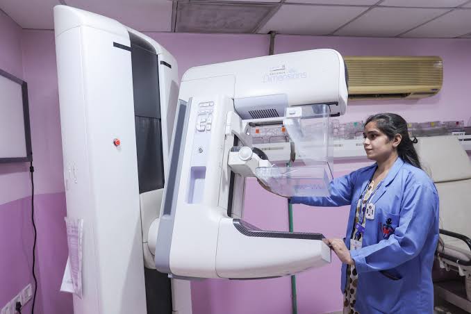
Digital Mammogram in Padur
Digital Mammography at AVR Scan Centre
A digital mammogram is a breast imaging examination that uses low-dose X-rays to create detailed images of breast tissue. It is commonly used as part of breast cancer screening and can also be recommended when a doctor needs to investigate a breast symptom or an abnormality found during another examination.
Digital mammography can help radiologists evaluate breast tissue for findings such as masses, calcifications and other changes that may require further assessment. The type and frequency of mammography recommended depend on factors such as age, symptoms, personal history and individual risk.
For patients looking for a Digital Mammogram in Padur, understanding why the examination has been recommended and what it can show can make the imaging process easier to approach.
Quick Answer – What Is a Digital Mammogram?
A digital mammogram is an X-ray-based breast imaging test that captures images of the breast electronically for examination by a radiologist. During the examination, the breast is positioned and briefly compressed between imaging surfaces so that clear images can be obtained.
Unlike a conventional film-based mammogram, digital mammography records the images electronically. The images can then be reviewed on a digital workstation and assessed for findings that may require further evaluation.
A digital mammogram may be performed for screening, when there are no known breast symptoms, or for diagnostic evaluation, when a patient has a symptom or a previously identified breast abnormality.
The appropriate type of mammographic examination depends on the individual's clinical situation. Your doctor or radiologist can determine which imaging approach is appropriate based on your symptoms, age, history and previous findings.
What Does a Digital Mammogram Show?
A digital mammogram can show changes in breast tissue that may not be detectable by physical examination alone. The images allow the radiologist to assess the appearance and distribution of breast tissue and identify areas that may need closer evaluation.
A mammogram can help demonstrate findings such as:
- Masses or areas of increased density that may require further assessment.
- Calcifications, which are small calcium deposits that can have different causes and appearances.
- Asymmetry, where one area of breast tissue appears different from the corresponding area.
- Architectural distortion, where the normal arrangement of breast tissue appears altered.
- Other changes that may warrant additional imaging or clinical correlation.
A finding on a mammogram does not automatically mean that cancer is present. Many breast findings are benign, while some require additional imaging or other evaluation to determine their significance.
The radiologist interprets the images together with the patient's clinical history and, when available, previous breast-imaging studies.
Why Is a Digital Mammogram Done?
A digital mammogram may be performed either for routine breast cancer screening or to evaluate a specific breast concern. The reason for the examination influences how the images are obtained and interpreted.
Screening Mammography
Screening mammography is performed in people who do not have a specific breast symptom but are undergoing recommended breast cancer screening.
The purpose is to look for certain breast changes that may warrant further assessment, including findings that can occur before a person notices a physical change.
Diagnostic Mammography
A diagnostic mammogram may be recommended when there is a particular concern, such as a breast lump, nipple change, focal breast pain or an abnormality identified during a previous examination or screening study.
In this situation, additional or targeted mammographic views may be obtained to evaluate the area more closely.
Screening and diagnostic mammography are therefore not interchangeable terms. The appropriate examination depends on why imaging has been recommended and what information the healthcare team needs.
Who May Need a Digital Mammogram?
People may be advised to undergo digital mammography as part of age- and risk-appropriate breast cancer screening or when a doctor needs to investigate a breast-related concern.
The decision to have a mammogram can depend on:
- Age and overall breast-cancer risk
- Personal history of breast conditions or cancer
- Family history and other risk factors
- A new breast lump or other physical change
- Nipple or skin changes
- An abnormal finding from a previous examination
- A recommendation following another breast-imaging test
There is no single screening schedule that is appropriate for every person. Different guidelines may recommend different approaches based on age and individual risk, so screening decisions should be discussed with an appropriate healthcare professional.
If you have a new or persistent breast symptom, do not wait for routine screening alone. A doctor may recommend diagnostic imaging or other evaluation based on the specific concern.
Digital Mammogram vs Traditional Mammogram – What Is the Difference?
The main difference is how the mammographic image is captured and stored. Traditional film mammography records the X-ray image on film, whereas digital mammography captures the image electronically.
Digital mammography allows breast images to be viewed and managed electronically, which can make image review and comparison with previous digital examinations more practical.
For the patient, however, the examination still involves breast positioning and compression during X-ray image acquisition. The clinical usefulness of the examination depends on obtaining appropriate images and having them accurately interpreted in the context of the patient's situation.
Digital mammography should therefore be understood as a modern method of acquiring and reviewing mammographic images, rather than assuming that every digital examination is automatically appropriate for every breast concern.
How Should You Prepare for a Digital Mammogram?
A digital mammogram usually requires very little preparation, and fasting is generally not required. You can usually eat and drink normally unless your healthcare provider has given you different instructions for another examination scheduled at the same visit.
On the day of your mammogram, it is helpful to:
- Wear clothing that allows you to undress from the waist up easily.
- Avoid applying deodorant, talcum powder, body lotion or other products to the breasts or underarms before the examination.
- Tell the imaging team if you are pregnant or think you could be pregnant.
- Inform the radiographer about breast implants, previous breast surgery or previous breast problems.
- Bring previous mammogram or breast-imaging reports and images when available.
If you have breast tenderness or are concerned about discomfort, you can mention this before the examination. The imaging professional can explain the positioning and compression process before beginning.
What Happens During a Digital Mammogram?
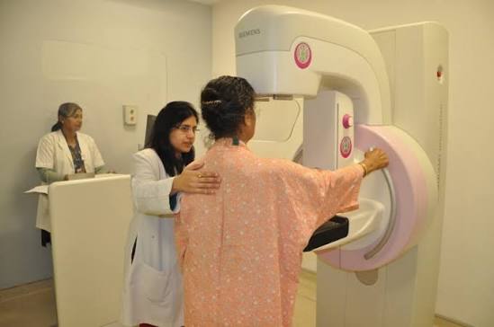
During a digital mammogram, each breast is positioned on the mammography equipment and briefly compressed while X-ray images are taken. The compression is an important part of obtaining useful images and helps spread the breast tissue for clearer evaluation.
The examination generally involves these steps:
- You will be asked to remove clothing from the upper body and any items that may interfere with the images.
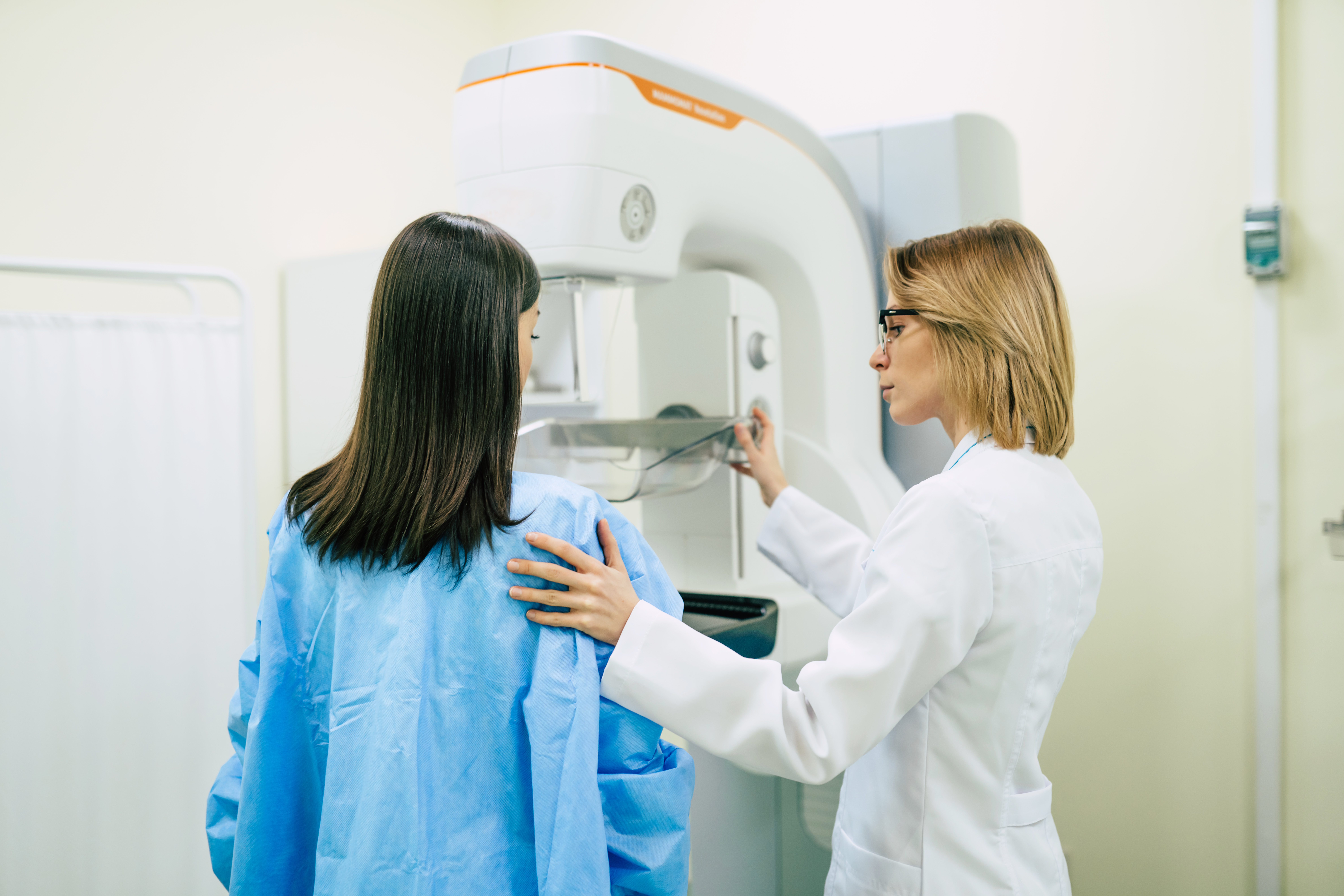
- The radiographer will position you in front of the mammography equipment.
- The breast will be placed on the imaging surface.
- A compression plate will gently but firmly compress the breast.
- The X-ray image will be obtained while you remain still.
- The compression is released after the image is captured.
- The process is repeated to obtain the required views of the breast.
More than one image of each breast may be required because different views allow the radiologist to assess breast tissue from different angles.
The radiographer may ask you to adjust your position or hold your breath briefly while an image is taken. These instructions help minimise movement and support the production of clear diagnostic images.
If additional images are required, this does not automatically mean that an abnormality has been found. Sometimes extra views are simply needed because the initial images do not provide enough information.
Does a Digital Mammogram Hurt?
A digital mammogram can cause temporary pressure or discomfort because the breast needs to be compressed during image acquisition. The experience varies between individuals, and some people may find the compression uncomfortable while others tolerate it easily.
The compression is brief and is released after each image is obtained. You can tell the radiographer if you are experiencing significant discomfort during the examination.
Breast tenderness can also vary during the menstrual cycle. If your breasts are particularly sensitive at certain times of the month, you may wish to discuss this when scheduling the examination.
It is important not to avoid a medically recommended mammogram solely because you are concerned about discomfort. If you are anxious about the procedure, letting the imaging professional know beforehand can help you understand what will happen and what to expect.
How Long Does a Digital Mammogram Take?
The actual mammographic image acquisition is usually relatively brief, but the total appointment time can vary depending on the examination and whether additional images are needed.
The overall visit may include:
- Registration and examination confirmation
- Changing into appropriate clothing
- Positioning for the mammogram
- Obtaining the required images
- Additional views when necessary
- Final completion of the imaging examination
A screening examination may follow a more straightforward process, while a diagnostic mammogram can involve additional targeted views depending on the reason for the examination.
Do not assume that needing additional images means that cancer has been detected. The radiologist may simply need more detailed views before completing the assessment.
If you have another appointment immediately afterward, it is sensible to allow some flexibility rather than planning around an exact examination time.
Can You Have a Digital Mammogram During Your Period?
Yes, a digital mammogram can generally be performed during your menstrual period, but breast tenderness may make the examination less comfortable for some people.
Hormonal changes can cause temporary breast swelling or sensitivity. If you have the flexibility to choose the appointment date and your breasts are usually very tender around menstruation, you can discuss suitable timing with the imaging centre.
However, a medically necessary diagnostic mammogram should not be unnecessarily delayed simply because you are menstruating. If your doctor has recommended imaging because of a breast lump, unusual change or another concern, follow the advised schedule unless your healthcare provider recommends otherwise.
If you are experiencing significant tenderness, inform the radiographer before the examination so they are aware of your discomfort.
When Will You Get Your Digital Mammogram Report?
The time required to receive a digital mammogram report depends on the examination, image review process and the diagnostic centre's reporting workflow.A screening mammogram and a diagnostic examination may involve different levels of image assessment and additional views.
The report is prepared after the mammographic images have been reviewed by the appropriate radiology professional. If additional images are needed, reporting may take longer because the radiologist may need to assess the additional views before completing the evaluation.
When booking your examination, it is useful to confirm:
- Expected report availability
- Whether the report will be provided digitally or in another format
- Whether previous images should be submitted for comparison
- How you will receive the report
- Whether a follow-up consultation is recommended
If you are waiting for a report after a screening mammogram, do not interpret the delay itself as an indication of an abnormal finding. Reporting time can vary for routine administrative and clinical reasons.
What Does a Digital Mammogram Report Tell You?
A digital mammogram report summarises the radiologist's assessment of the breast images and indicates whether any finding requires further evaluation or routine follow-up.
Depending on the examination and reporting system, the report may describe:
- The overall appearance of the breast tissue
- Any masses, calcifications or areas of asymmetry
- Changes compared with previous mammograms, when available
- Breast density where it is assessed and reported
- Whether additional imaging or follow-up is recommended
- An overall imaging assessment or category
One important point is that a mammogram report should not be interpreted by focusing on a single word or finding in isolation. The radiologist considers the appearance of the breast tissue, previous imaging and the clinical reason for the examination.
If the report recommends additional imaging, this does not automatically mean that cancer has been diagnosed. Additional views, ultrasound or other assessment may be recommended simply to clarify an imaging finding.
If you are unsure about a result, discuss the complete report with your doctor or the appropriate breast-imaging specialist.
Is a Digital Mammogram Safe?
Digital mammography uses X-rays and therefore involves a small amount of radiation exposure. Mammography equipment is designed to use a low radiation dose while obtaining images suitable for breast evaluation.
For people undergoing recommended mammography, the potential benefits of appropriate breast screening or diagnostic assessment are considered in relation to this small radiation exposure.
Before the examination, tell the imaging team if you are pregnant or think you may be pregnant. This information is important when deciding whether mammography is appropriate and how the examination should be managed.
The benefits and suitability of mammography can also differ depending on the reason for the examination. A screening examination and an examination performed to investigate a concerning symptom are different clinical situations.
If you have concerns about radiation exposure, pregnancy or repeated mammograms, discuss them with your doctor or radiology team rather than avoiding medically recommended imaging.
Can a Digital Mammogram Detect Breast Cancer?
A digital mammogram can help detect certain breast abnormalities associated with breast cancer, including some cancers that may not yet cause noticeable symptoms.It is an important imaging tool for breast cancer screening and diagnostic assessment.
Mammography can identify changes such as suspicious masses, calcifications or architectural distortion that may require further evaluation. However, no imaging test can guarantee detection of every breast cancer.
The ability of mammography to show an abnormality can be influenced by factors such as breast density, the characteristics of the lesion and the type of cancer involved.
If a mammogram identifies a suspicious finding, additional assessment may be recommended. This can include additional mammographic views, ultrasound, breast MRI in selected circumstances or tissue sampling when clinically indicated.
Conversely, if a mammogram is reported as normal, you should still seek medical assessment for a new or persistent breast symptom. A normal mammogram should not be used to dismiss a concerning clinical finding.
When Is an Ultrasound Needed Along With a Mammogram?
A breast ultrasound may be recommended alongside mammography when additional information is needed to evaluate a particular finding or clinical concern.. Mammography and ultrasound use different imaging methods and can provide complementary information.
An ultrasound may be considered in situations such as:
- Further evaluation of a breast lump
- Clarifying a finding seen on mammography
- Assessing a particular area of breast tissue
- Evaluating certain findings in dense breast tissue
- Distinguishing some fluid-filled structures from solid ones
However, ultrasound is not automatically required for everyone who has a mammogram. Whether it is useful depends on the mammographic findings, breast characteristics, symptoms and clinical question.
If additional ultrasound is recommended after your mammogram, it does not necessarily mean that cancer is suspected. The purpose may simply be to obtain more information about an area seen on the initial examination.
How Much Does a Digital Mammogram Cost in Padur?
The cost of a digital mammogram in Padur depends on the type of examination and the services included by the diagnostic centre. A routine screening examination may have different charges from a diagnostic examination requiring additional views or complementary imaging.
Before booking, confirm:
- Current mammogram price
- Whether the quoted charge covers the required views
- Whether additional views are charged separately
- Whether a breast ultrasound is included or booked separately
- Report availability
- Appointment requirements
For the current Digital Mammogram price at AVR Scan Centre, contact the centre directly for the latest details. Prices and service arrangements can change, so confirming the current charge is preferable to relying on an outdated online figure.
If your doctor has prescribed a particular mammographic examination, provide the prescription or referral details when enquiring so the centre can confirm the appropriate service and applicable charge.
How Should You Choose a Digital Mammogram Centre in Padur?
When choosing a centre for a digital mammogram in Padur, consider the quality of the imaging service, availability of appropriate mammography facilities and the clarity of the reporting process rather than choosing only on price.
Before booking, it is useful to confirm:
- Whether digital mammography is available for your specific requirement
- Whether screening or diagnostic mammography is appropriate
- Whether previous mammograms can be provided for comparison
- How the images and report will be reviewed
- Whether additional views can be arranged if required
- Current charges and appointment availability
- Whether the centre provides clear preparation instructions
For patients who have been referred for a particular breast-imaging examination, sharing the doctor's prescription or referral details when booking can help the centre understand the reason for the examination.
Convenient location can also matter because breast imaging sometimes involves additional views or follow-up investigations. A centre in or around Padur may be practical for patients who need to return for further evaluation.
What Should You Bring for a Digital Mammogram?
Bringing relevant medical and imaging information can help the radiology team understand your current examination and compare it with previous findings when necessary.
Consider bringing:
- Your doctor's prescription or referral, if provided
- Previous mammogram reports
- Previous mammogram images, particularly if performed at another centre
- Relevant breast-ultrasound or other breast-imaging reports
- Details of previous breast surgery or procedures
- Information about breast implants, if applicable
- A list of relevant medicines or medical history when requested
Previous images can be particularly useful because comparison with earlier examinations may help the radiologist identify whether a finding is new, unchanged or has developed over time.
If you have noticed a recent breast lump, nipple change, skin change or another concern, mention it to the healthcare team rather than relying only on the written referral.
What Happens After a Digital Mammogram?
After the mammographic images are obtained, they are reviewed by the radiology team and a report is prepared based on the findings. The next step depends on the result and the reason the examination was performed.
Possible outcomes include:
- Routine screening follow-up when no concerning finding is identified
- Comparison with previous mammograms
- Additional mammographic views
- Breast ultrasound or another imaging examination
- Clinical review by the referring doctor
- Further investigation when a suspicious finding requires clarification
A recommendation for additional imaging does not automatically mean that cancer is present.
A recommendation for additional imaging does not automatically mean that cancer is present. It may simply mean that the radiologist needs more information before reaching a final assessment.
If a biopsy or other investigation is recommended, the decision should be discussed with the appropriate doctor or breast specialist. The mammogram itself is an imaging examination and does not by itself establish a final tissue diagnosis.
Why Choose AVR Scan Centre for Digital Mammography in Padur?
AVR Scan Centre provides diagnostic imaging services for patients seeking breast imaging in the Padur and OMR area. Patients who have been advised to undergo digital mammography can contact the centre to confirm the appropriate examination and current appointment details.
Patients can confirm:
- Availability of digital mammography
- Whether the examination is intended for screening or diagnostic evaluation
- Current charges
- Appointment availability
- Preparation instructions
- Requirements for bringing previous breast-imaging records
- Report availability and collection details
If you have a doctor's prescription or referral, provide the exact details when contacting the centre so the appropriate examination can be confirmed.
For patients in Padur who need breast imaging, arranging the examination at a conveniently located diagnostic centre can also make subsequent imaging or follow-up easier when recommended.
Digital Mammogram – Frequently Asked Questions
Yes, women with breast implants can undergo mammography, but the imaging team should be informed about the implants before the examination. Special positioning techniques may be used to obtain useful images of the breast tissue while taking the implants into account.
Mammography may be performed after breast surgery when clinically appropriate, but the timing and type of imaging depend on the surgery and reason for the examination. Tell the imaging team about previous breast surgery so the radiologist can interpret the images with the relevant history.
It is generally advisable to avoid deodorant, talcum powder, lotion or similar products on the breasts and underarms before a mammogram. Some products can contain particles that may appear on the X-ray images and potentially make interpretation more difficult.
Yes, previous mammogram reports and images can be valuable for comparison. If your earlier examination was performed elsewhere, ask whether the previous images can be provided in an appropriate format and bring them along with the report.
Mammography can sometimes be performed during breastfeeding when there is a clinical reason to investigate a breast concern. Tell the imaging team that you are breastfeeding so the examination can be considered in the appropriate clinical context. Depending on the situation, other imaging may also be recommended.
Yes, mammography can be performed in men when a doctor considers breast imaging necessary. It may be recommended to investigate a lump, nipple change, breast enlargement or another concerning finding.
No. A mammogram uses X-rays to create breast images, while breast ultrasound uses sound waves. They provide different types of information and may sometimes be used together depending on the patient's symptoms, breast characteristics and mammographic findings.
An abnormal mammogram does not automatically mean that you have breast cancer. The radiologist may recommend additional mammographic views, ultrasound, clinical assessment or another investigation to clarify the finding. Follow-up should be based on the complete imaging report and your doctor's advice.
Book a Digital Mammogram in Padur
If you have been advised to undergo digital mammography in Padur, confirm the examination details before visiting the centre.
Before booking, check:
- The type of mammogram required
- Current charges
- Appointment availability
- Preparation instructions
- Whether previous mammogram images and reports should be brought
- Expected report availability
- Whether additional breast imaging can be arranged if recommended
Contact AVR Scan Centre to confirm current digital mammography availability, preparation requirements, pricing and appointment details.
AVR Scan Centre – Padur
Godrej Azure Elsa Plaza, #3, Ground Floor, Padur, Tamil Nadu – 603103.
