
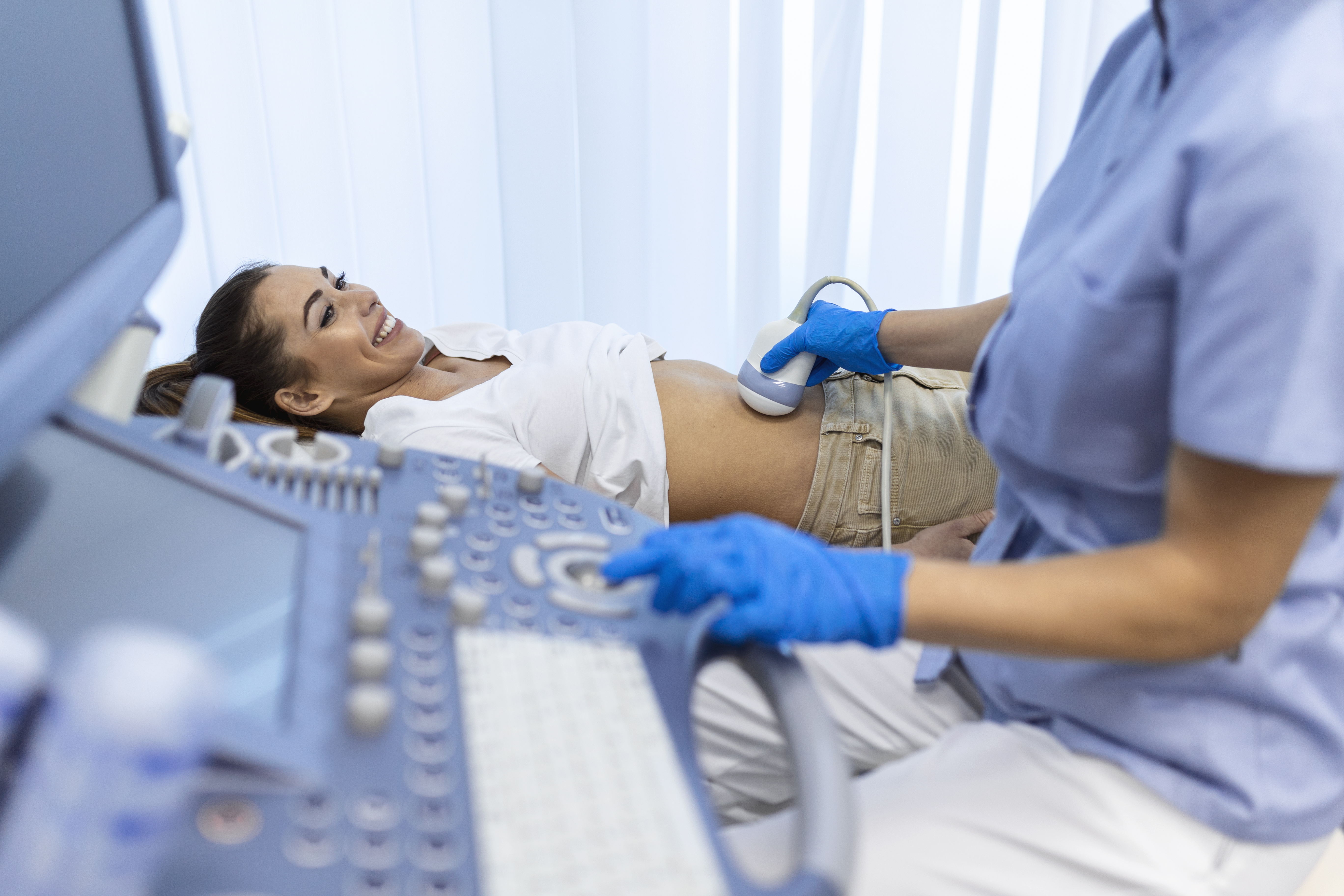
Ultrasound Scan in Padur
Diagnostic Ultrasound at AVR Scan Centre
If your doctor has recommended an ultrasound, you may be looking for an Ultrasound Scan in Padur and want to know which type of scan you may need, whether preparation is required and what the examination involves.
Ultrasound, also called ultrasonography or sonography, uses high-frequency sound waves to create images of structures inside the body. It can provide real-time imaging and is commonly used to examine selected organs, soft tissues and pregnancy-related structures.
AVR Scan Centre provides Ultrasound Scan services in Padur for patients looking for diagnostic imaging in and around Padur and the OMR region.
The right ultrasound depends on the body area being examined, your symptoms, medical history and the clinical question your doctor needs to answer. Preparation also varies by examination, so fasting or a full bladder may be needed for some scans but not others.
Is an Ultrasound Scan Safe?
Ultrasound does not use ionizing radiation and is widely used for diagnostic imaging when medically appropriate. It uses sound waves to create images rather than X-rays.
Ultrasound may be used for many different examinations, including selected abdominal, pelvic and pregnancy-related scans.
The appropriate scan should be based on the clinical need and performed according to the instructions given by your healthcare professional.
Ultrasound Scan in Padur – Location, Availability & Appointment
AVR Scan Centre provides ultrasound services at its Padur centre for patients looking for diagnostic imaging in and around Padur.
AVR Scan Centre – Padur
Godrej Azure Elsa Plaza, #3, Ground Floor, Padur, Tamil Nadu – 603103.
The Padur centre is relevant for patients searching for an Ultrasound Centre in Padur or an Ultrasound Scan near OMR.
Before visiting, confirm:
- The exact ultrasound examination required
- Whether preparation is needed
- Whether fasting is required
- Whether a full bladder is needed
- Appointment availability
- Any specific instructions for your examination
What Can an Ultrasound Scan Show?
An ultrasound can produce real-time images of selected organs, tissues and other structures inside the body.
Depending on the examination, ultrasound may help evaluate areas such as:
- Abdomen
- Pelvis
- Urinary system
- Selected soft tissues
- Selected organs
- Pregnancy-related structures
Ultrasound can also be used to assess movement or blood flow in certain examinations when Doppler techniques are included.
The information available from ultrasound depends on the body part being examined and the type of scan performed.
Which Ultrasound Scan Do You Need?
The appropriate ultrasound examination depends on the body area, symptoms and the reason your doctor has requested imaging.
Abdominal Ultrasound
An abdominal ultrasound can be used to assess selected organs and structures within the abdomen, depending on the clinical question.
Pelvic Ultrasound
A pelvic ultrasound is used to examine structures within the pelvis and may be recommended for selected symptoms or clinical concerns.
Pregnancy or Obstetric Ultrasound
Ultrasound is commonly used during pregnancy to assess pregnancy-related structures and development according to the stage of pregnancy and the clinical requirement.
Urinary-System Ultrasound
Ultrasound may be used to assess selected structures of the urinary system when clinically indicated.
Other Region-Specific Ultrasound Examinations
Other ultrasound examinations may be performed depending on the area being assessed and the service available.
Do not choose an ultrasound type solely from an online search. The examination should match your doctor's recommendation.
Why Does Your Doctor Recommend an Ultrasound?
A doctor may recommend an ultrasound when real-time imaging can help evaluate a symptom, body structure or suspected condition.
Ultrasound may be considered when a doctor needs information about:
- Abdominal or pelvic symptoms
- Selected urinary concerns
- Certain soft-tissue problems
- Pregnancy-related assessment
- Selected organ-related findings
- Other conditions where ultrasound is clinically useful
The choice depends on:
- Symptoms
- Medical history
- Physical examination
- Previous investigations
- The body area requiring evaluation
Ultrasound is one imaging option among several, and the most suitable test depends on the clinical question.
Why Do Some Ultrasound Scans Need Fasting or a Full Bladder?
Preparation depends on the type of ultrasound being performed. Some examinations may require fasting, while others may require a comfortably full bladder to improve the view of certain structures.
The reason for preparation can depend on:
- Body area being examined
- Type of ultrasound
- Doctor's clinical question
- Imaging protocol
Do not assume that every ultrasound has the same preparation requirements.
Do You Need to Fast Before an Ultrasound?
Not every ultrasound requires fasting. Some abdominal examinations may involve fasting, while many other ultrasound scans do not.
If fasting is required, the centre should provide the appropriate instructions for your examination.
Before your appointment, confirm:
- How long you need to fast
- Whether plain water is allowed
- Whether regular medicines should be taken
- Whether any other preparation is needed
Follow the instructions for your specific scan rather than general fasting advice.
Should You Drink Water Before an Ultrasound?
Some ultrasound examinations may require you to drink water and have a full bladder, while others do not.
A full bladder may help improve the imaging of selected pelvic or urinary structures.
Your preparation can therefore vary depending on the examination. Confirm with the centre whether you should:
- Drink a specified amount of water
- Avoid emptying your bladder before the scan
- Follow any additional preparation instructions
What Should You Bring for an Ultrasound Appointment?
For your ultrasound visit, it can be useful to carry:
- Doctor's prescription or referral
- Previous ultrasound or other imaging reports
- Relevant medical records
- Details of current medicines
- Any test-specific documents requested by the centre
Previous reports may help your doctor compare current findings with earlier examinations when appropriate.
What Happens During an Ultrasound Scan?
During an ultrasound examination, you are usually positioned so the area being examined can be accessed clearly.
The usual process may include:
- Confirming the requested ultrasound examination.
- Positioning you appropriately.
- Applying ultrasound gel to the skin.
- Moving a handheld device called a transducer over the area being examined.
- Capturing images in real time.
- Taking additional views when needed.
Depending on the examination, you may be asked to:
- Remain still
- Change position
- Hold your breath briefly
- Follow other simple instructions
The exact process depends on the body area and type of ultrasound.
Is an Ultrasound Scan Painful?
Ultrasound is generally non-invasive and is usually not painful.
You may feel some pressure when the transducer is moved over the skin. This can be more noticeable when the area being examined is already tender or painful.
Tell the healthcare professional if you experience significant discomfort during the examination.
Is Ultrasound Safe During Pregnancy?
Ultrasound does not use ionizing radiation and is widely used for pregnancy-related imaging when clinically indicated.
The type and timing of an obstetric ultrasound depend on:
- Stage of pregnancy
- Clinical requirement
- Doctor's recommendation
- Findings from previous examinations
Pregnancy ultrasound should be performed for an appropriate medical or obstetric purpose rather than simply because a scan is available.
What Is a Doppler Ultrasound?
Doppler ultrasound is a specialised form of ultrasound that can assess the movement of blood through certain blood vessels.
It uses changes in reflected sound waves to provide information about blood flow.
A doctor may recommend Doppler imaging when information about blood movement is important for the clinical question being investigated.
Doppler techniques can be used in selected:
- Blood vessel examinations
- Pregnancy-related assessments
- Other studies where blood-flow information is clinically relevant
Doppler is not required for every ultrasound scan. It is used when blood-flow information adds value to the examination.
What Is the Difference Between Ultrasound and Doppler?
A standard ultrasound mainly creates images of anatomical structures, while Doppler ultrasound adds information about blood flow.
| Standard Ultrasound | Doppler Ultrasound |
|---|---|
| Produces images of structures | Can also assess blood flow |
| Commonly used for organs and soft tissues | Used when blood-flow information is needed |
| May be sufficient for many examinations | Often added to selected examinations |
| Does not always require Doppler information | Provides additional flow-related information |
The appropriate examination depends on what your doctor needs to assess.


Can Ultrasound Find Every Problem?
No. Ultrasound is useful for many conditions, but it cannot detect every disease or provide the same information as CT, MRI or other diagnostic tests.
Its usefulness can depend on:
- Body area
- Type of condition
- Patient-related factors
- Depth and location of the structure
- The type of ultrasound performed
In some situations, your doctor may recommend another investigation when ultrasound does not provide enough information.
A normal ultrasound does not necessarily rule out every possible cause of a symptom.
Ultrasound vs CT vs MRI – Which Test Is Used for What?
Ultrasound, CT and MRI use different technologies and are chosen for different clinical needs.
| Ultrasound | CT | MRI |
|---|---|---|
| Uses sound waves | Uses X-rays | Uses magnetic fields and radio waves |
| Does not use ionizing radiation | Uses ionizing radiation | Does not use ionizing radiation |
| Useful for many organs, soft tissues and pregnancy-related imaging | Provides detailed cross-sectional imaging | Useful for many detailed soft-tissue examinations |
| Can provide real-time imaging | Can provide rapid cross-sectional images | Often provides detailed images in multiple planes |
Which One Is Better?
There is no single imaging test that is best for every condition.
Your doctor selects the examination based on:
- Symptoms
- Body area
- Suspected condition
- Medical history
- Type of information required
How Long Does an Ultrasound Scan Take?
The duration of an ultrasound depends on the body area, type of examination and preparation required.
The appointment may take longer when:
- Multiple areas are examined
- Additional views are needed
- Doppler assessment is included
- Patient positioning needs to be changed
- The examination requires specific preparation
For the expected duration of your particular ultrasound in Padur, confirm the details when booking.
How Much Does an Ultrasound Scan Cost in Padur?
The cost of an ultrasound in Padur can vary depending on the type of examination and whether any specialised imaging, such as Doppler, is included.
Before booking, confirm:
- Exact ultrasound examination
- Whether Doppler is included
- Current scan charges
- Any additional requirements
For the current price at AVR Scan Centre, contact the Padur centre rather than relying on an outdated online price.
What Should You Check Before Booking an Ultrasound in Padur?
Before confirming your appointment, check:
- The exact ultrasound prescribed
- Whether fasting is required
- Whether a full bladder is needed
- Whether Doppler is included
- Whether pregnancy-related information is relevant
- Whether previous reports should be brought
- Appointment availability
- Current pricing
The preparation should match the exact ultrasound examination rather than a general ultrasound instruction.
What Happens After an Ultrasound Scan?
After the ultrasound examination, the images are reviewed and a report is prepared based on the findings and the type of examination performed.
The result may be considered along with:
- Your symptoms
- Medical history
- Physical examination
- Previous imaging
- Other test results
If the findings need further evaluation, your doctor may recommend another investigation, follow-up imaging or additional clinical assessment.
An ultrasound report should be interpreted together with your overall clinical information rather than from one finding alone.
How Are Ultrasound Findings Reported?
An ultrasound report describes the findings seen during the examination and may include measurements, observations and conclusions relevant to the scan performed.
The report may contain:
- Examination name
- Structures examined
- Measurements, where relevant
- Observed findings
- Impression or conclusion
The wording and level of detail can vary depending on the type of ultrasound.
If you are unsure about a finding in the report, ask your doctor to explain what it means in relation to your symptoms and medical history.
Why Choose AVR Scan Centre for an Ultrasound in Padur?
When choosing an Ultrasound Centre in Padur, patients may consider the location, availability of the required examination and how the centre handles preparation and appointment requirements.
AVR Scan Centre provides Ultrasound Scan services in Padur for patients seeking diagnostic imaging in and around Padur and the OMR region.
The centre also provides other diagnostic services, which may be useful when additional investigations are recommended by a doctor.
Only verified AVR-specific information should be added for claims related to:
- Specialist credentials
- Accreditation
- Reporting timelines
- Special ultrasound equipment
- Home services
- Package benefits
- Pricing
Ultrasound Scan in Padur – Questions People Often Ask
A repeat ultrasound may be appropriate when your doctor needs follow-up information or wants to compare findings over time.
Yes. Previous reports can provide useful context and may help compare current findings with earlier examinations.
Ultrasound may be used in children when clinically appropriate. The type of examination depends on the child's symptoms and the clinical question.
The number and timing of pregnancy ultrasounds depend on the pregnancy, medical need and the recommendation of the treating healthcare professional.
Ultrasound can detect certain cysts, stones and other findings in appropriate body areas, but its ability depends on the location, size and type of abnormality.
Ultrasound can provide information about the appearance of the liver, kidneys and other abdominal structures. The significance of a finding depends on the complete clinical context.
This can depend on the examination and centre procedures. Confirm with the centre before your appointment if another person needs to accompany you.
A routine external ultrasound generally does not require recovery time. Follow any specific instructions given for your examination.
Get Your Ultrasound Scan at AVR Scan Centre, Padur
If your doctor has recommended an ultrasound, knowing the exact examination, preparation requirements and appointment process can help you prepare for the visit.
AVR Scan Centre provides Ultrasound Scan services in Padur for patients looking for diagnostic imaging in and around the OMR region.
Before visiting, confirm:
- The exact ultrasound examination
- Preparation requirements
- Whether fasting or a full bladder is needed
- Appointment availability
- Current scan pricing
Need an Ultrasound Scan in Padur?
Contact AVR Scan Centre – Padur to enquire about the ultrasound examination recommended by your doctor and confirm the requirements for your appointment.
Contact AVR Scan Centre – Padur to enquire about the ultrasound examination, preparation requirements and appointment availability.
AVR Scan Centre – Padur
Godrej Azure Elsa Plaza, #3, Ground Floor,
Padur, Tamil Nadu – 603103.
