
SonoMammography in Padur
Breast Ultrasound at AVR Scan Centre
If you have been advised to undergo SonoMammography in Padur, you may want to understand what the examination is used for, what it can show and how it differs from other breast-imaging tests.
SonoMammography, commonly referring to breast ultrasound, uses sound waves to create images of breast tissue. It can be used to examine a specific area of concern, such as a breast lump or other change, and may provide additional information when another breast examination needs further assessment.
AVR Scan Centre provides SonoMammography services in Padur for patients looking for breast imaging in and around Padur and the OMR region.
The appropriate breast-imaging test depends on the reason for the examination and factors such as symptoms, age, medical history and the findings that need to be assessed. Breast ultrasound is useful in selected situations, but it does not automatically replace mammography or other breast investigations.
Quick Answer – What Is SonoMammography?
SonoMammography is an ultrasound examination of the breast that uses sound waves to create images of breast tissue. It does not use ionizing radiation and can help assess certain breast lumps, cysts and other areas that need closer examination.
It may be performed as a focused examination of a particular breast concern or as an additional imaging test when more information is needed.
The findings should be interpreted in the context of the patient's symptoms, clinical examination and other breast-imaging results when relevant.
Where Can You Get SonoMammography in Padur?
AVR Scan Centre provides SonoMammography in Padur for patients looking for breast ultrasound services in the Padur and OMR region.
AVR Scan Centre – Padur
Godrej Azure Elsa Plaza, #3, Ground Floor, Padur, Tamil Nadu – 603103.
The service may be relevant for people searching for:
- SonoMammography in Padur
- Breast Ultrasound in Padur
- Breast Sonography in Padur
- Breast Scan Centre in Padur
- Breast Ultrasound near OMR
If your doctor has recommended a specific breast examination, confirm the exact scan, appointment availability and any centre-specific instructions before visiting.
If you have previous mammograms, breast ultrasounds or other relevant reports, it is also useful to ask whether you should bring them for comparison.
When Is SonoMammography Used for Breast Problems?
Breast ultrasound may be recommended when a doctor needs more information about a specific breast concern or wants to examine an area more closely.
Depending on the clinical situation, a doctor may recommend it when there is:
- A new or palpable breast lump
- Localised breast pain
- A breast change noticed during a physical examination
- An area that needs further assessment after another imaging test
- A breast finding that needs additional characterisation
- A breast concern in a younger patient where ultrasound may provide useful information
- A breast concern during pregnancy when breast imaging is clinically appropriate
The reason for the examination is important. A diagnostic breast ultrasound for a specific concern is different from choosing an imaging test simply as a general health check.
Depending on your age, symptoms, medical history and the finding being investigated, your doctor may recommend ultrasound alone or combine it with another breast-imaging examination.
What Information Can Breast Ultrasound Provide About a Finding?
Breast ultrasound can provide detailed images of a specific area and help describe the appearance of a breast finding.
For example, ultrasound may help assess whether an area appears:
- Fluid-filled
- Solid
- Well-defined or less clearly defined
- Different from the surrounding breast tissue
- Suitable for follow-up or further assessment
This can be useful when a patient or doctor has identified a particular area that needs evaluation. Ultrasound can provide additional information about the nature of a finding and may help determine whether further assessment is appropriate.
However, an ultrasound image by itself does not establish a diagnosis. A breast finding may be benign, require monitoring or need additional investigation depending on its appearance and the patient's clinical history.
If a finding requires further assessment, the next step may involve another imaging examination or, when clinically indicated, tissue sampling.
The purpose of the examination is therefore not simply to label an image as “normal” or “abnormal.” The findings need to be interpreted as part of the patient's overall breast assessment.
How Should You Prepare for a SonoMammography Scan?
SonoMammography usually requires very little preparation, and most patients can follow their normal routine before the examination. Unlike some medical tests, a breast ultrasound generally does not require fasting or special dietary preparation.
Before your appointment, it is useful to:
- Wear comfortable clothing that can be removed easily from the upper body.
- Avoid applying thick creams, lotions or other products over the breast area immediately before the scan.
- Bring previous mammography or breast ultrasound reports if they are available.
- Carry your doctor's prescription or referral when applicable.
- Inform the healthcare professional about any relevant breast symptoms or medical history.
If you have noticed a particular lump, painful area or other change, tell the healthcare professional where it is located. This information can help ensure that the area of concern receives appropriate attention during the examination.
Preparation can vary if the breast ultrasound is being performed along with another examination. Follow any specific instructions provided by the centre or your doctor rather than assuming that every breast-imaging appointment has identical requirements.
What Happens During a Breast Ultrasound?
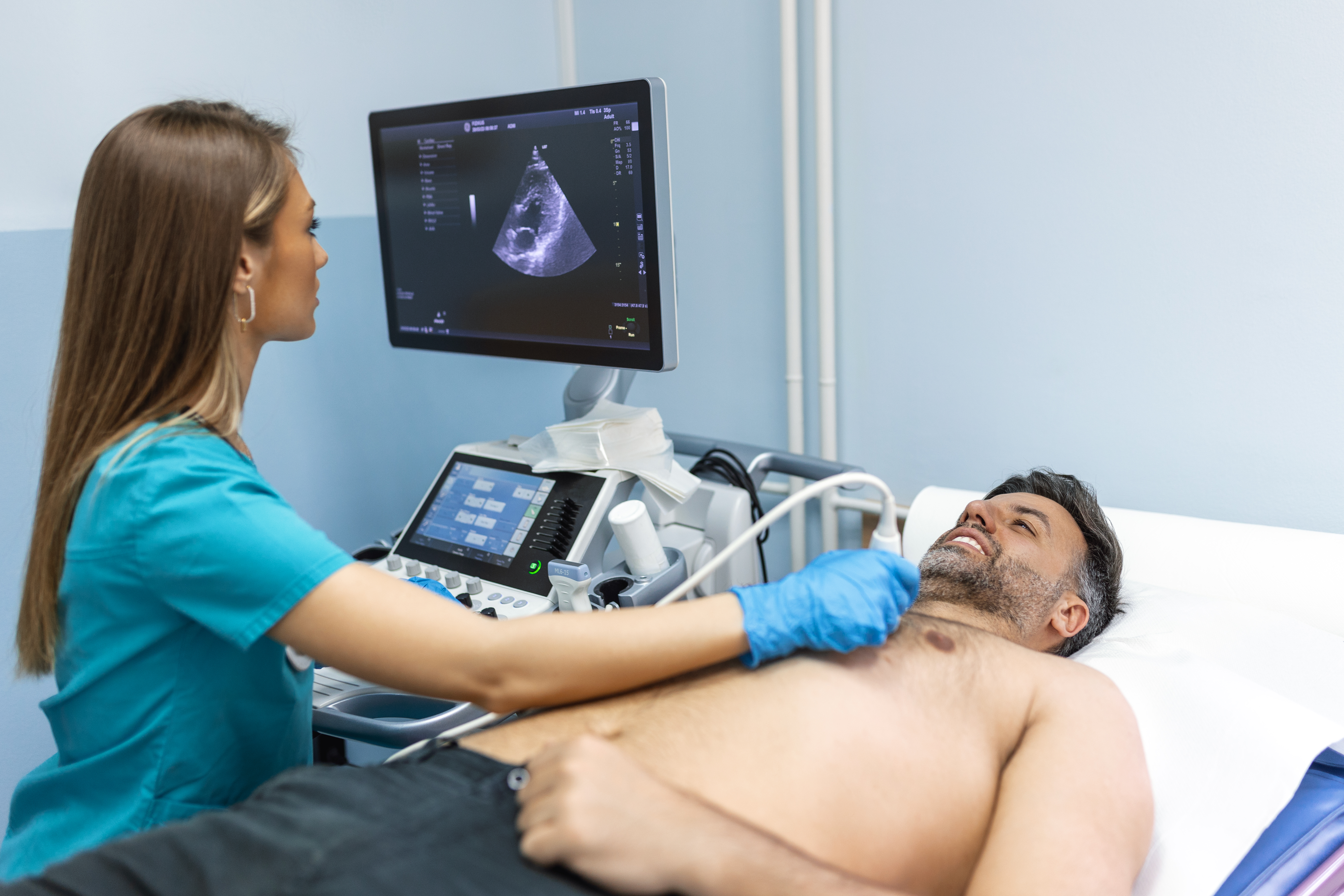
During a breast ultrasound, you will usually lie in a comfortable position while an ultrasound transducer is moved over the breast. A clear ultrasound gel is applied to the skin to help the transducer move smoothly and allow the sound waves to travel effectively through the tissues.
The examination generally involves:
- You will be asked to lie in a suitable position.
- The breast area being examined will be exposed while maintaining appropriate privacy.
- Ultrasound gel will be applied to the skin.
- The transducer will be moved gently across the breast.
- Images will be captured from different angles as needed.
- Additional views may be taken if a particular area needs closer assessment.
- The images will be reviewed and used to prepare the examination report.
If the scan was requested because of a lump, pain or another specific concern, tell the healthcare professional before the examination. Knowing the location of the concern can help direct the examination to the relevant area.
Depending on the clinical reason for the scan, the area around the armpit may also be examined when appropriate.
The scan does not involve needles or injections as part of a standard ultrasound examination.
Is SonoMammography Painful?
Breast ultrasound is generally non-invasive and does not usually cause significant pain. The examination is performed by moving the ultrasound transducer over the skin with the help of gel.
You may feel some pressure while the transducer is moved across the breast. This pressure can feel uncomfortable if you already have a tender lump, breast pain or another sensitive area.
You should tell the healthcare professional if a particular area is painful. This allows the examination to be performed as comfortably as possible while still obtaining the necessary images.
The scan itself does not involve radiation, needles or an incision.
Is Breast Ultrasound Safe During Pregnancy?
Breast ultrasound does not use ionizing radiation and may be used during pregnancy when breast imaging is clinically needed.
Pregnancy can cause natural changes in breast tissue. Because of these changes, a new lump or other breast concern may sometimes need imaging rather than physical examination alone.
If you are pregnant or think you may be pregnant, tell your doctor and the imaging team before the examination. They can determine the most appropriate imaging approach based on the nature of the breast concern and your individual situation.
A new or persistent breast change should not be ignored simply because you are pregnant. If you notice a lump, unusual discharge, skin change or another breast concern, discuss it with your healthcare professional.
Is SonoMammography the Same as a Mammogram?
No. SonoMammography and mammography are different breast-imaging tests that use different methods and provide different types of information.
SonoMammography, or breast ultrasound, uses sound waves to create images of breast tissue. It can be useful when a doctor needs to examine a particular area, such as a lump, or wants additional information about a breast finding.
A mammogram uses low-dose X-rays to create images of the breast. Mammography has an important role in breast cancer screening and can show certain findings, including some calcifications, that may not be assessed as well with ultrasound.
The main difference can be understood simply:
| SonoMammography / Breast Ultrasound | Mammography |
|---|---|
| Uses sound waves | Uses low-dose X-rays |
| Does not use ionizing radiation | Uses ionizing radiation |
| Useful for assessing selected breast findings | Important for breast cancer screening |
| Can provide information about certain cystic and solid-appearing findings | Can show certain calcifications and other abnormalities |
Neither examination is automatically better for every patient. Your doctor may recommend ultrasound, mammography or both depending on your age, symptoms, breast tissue, medical history and the reason for the examination.
Can SonoMammography Detect Breast Cancer?
Breast ultrasound can identify some suspicious breast abnormalities, but it cannot detect every breast cancer or confirm cancer by itself.
Ultrasound can provide useful information about the appearance of a breast finding and may help identify an area that needs further evaluation. It can be particularly useful when a specific lump or area needs closer assessment.
However, some breast cancers may not be clearly visible on ultrasound. This is one reason ultrasound should not automatically be considered a substitute for mammography when mammography is appropriate.
If an ultrasound finding needs further evaluation, your doctor may recommend:
- Additional mammography
- Follow-up breast imaging
- Another imaging examination in selected situations
- Tissue sampling or biopsy when clinically required
A suspicious ultrasound finding does not automatically mean that cancer is present. At the same time, a normal ultrasound does not guarantee that every breast abnormality has been excluded.
The result should therefore be considered together with your symptoms, physical examination, previous imaging and medical history.
Is Breast Ultrasound Useful for Dense Breasts?
Breast ultrasound can provide additional information in some patients with dense breast tissue, but having dense breasts does not automatically mean that everyone needs an ultrasound.
Breast density refers to the relative amount of fibroglandular tissue in the breast. Dense tissue can make some abnormalities harder to see clearly on a mammogram.
In selected situations, ultrasound may be used as an additional examination to assess areas that need further evaluation. It can provide information that complements what is seen on mammography.
Whether additional ultrasound is appropriate depends on factors such as:
- Breast density
- Age
- Personal medical history
- Family history
- Individual breast cancer risk
- Mammography findings
- Current symptoms
The decision should be based on your individual risk and clinical situation, not on breast density alone.
What Happens If a Breast Ultrasound Finds a Lump?
A lump seen on breast ultrasound does not automatically mean that it is cancer. Breast lumps can have different causes, including benign cysts and other non-cancerous conditions.
Ultrasound can help describe the appearance of a lump and provide information that may guide the next step. Depending on the findings, your doctor may recommend routine follow-up, additional imaging or tissue sampling.
The next step can depend on:
- The appearance of the finding
- Its size and location
- Whether it appears fluid-filled or solid
- Your age and medical history
- Whether the lump can be felt clinically
- Findings from other breast examinations
If a biopsy is recommended, this does not mean that cancer has already been confirmed. A biopsy is used to obtain tissue when imaging alone cannot provide a definite answer.
A new or persistent breast lump should be assessed by a healthcare professional rather than judged from an ultrasound image alone.
How Much Does SonoMammography Cost in Padur?
The cost of SonoMammography in Padur depends on the diagnostic centre and the specific examination being performed.
Before booking, confirm the current price for the breast ultrasound recommended for you. It can also be useful to clarify whether the examination is focused on one area or involves a broader assessment.
Ask about:
- Current scan charges
- The exact examination included
- Whether additional imaging has been recommended
- Appointment availability
- Any preparation or documents required
For the current SonoMammography price at AVR Scan Centre, contact the Padur centre directly rather than relying on an older online price.

What Should You Check Before Booking SonoMammography in Padur?
Before arranging an appointment, first confirm the exact breast-imaging examination your doctor has recommended and the reason it is being performed.
It is useful to have:
- Your prescription or referral, where applicable
- Previous mammography reports
- Previous breast ultrasound reports
- Relevant medical history
- Information about a particular lump, pain or other breast change
- Pregnancy information, when relevant
You can also confirm the current appointment availability and examination charge before visiting.
If you have already had breast imaging elsewhere, ask whether bringing the previous reports or images would be useful for comparison. This can help the healthcare professional consider the current findings alongside earlier information.
What Happens After a SonoMammography Scan?
After the breast ultrasound is completed, the captured images are reviewed and the findings are documented in a report. The report describes the areas examined and any relevant features seen during the scan.
The findings may be considered together with:
- The reason for the examination
- Your current symptoms
- Clinical breast examination
- Previous ultrasound or mammography results
- Relevant medical history
The outcome can vary depending on what was seen. Some findings may simply require routine follow-up, while others may need additional imaging or further clinical assessment.
If the scan was performed to investigate a particular lump or other breast change, the report may describe the location and appearance of that finding.
The next step should be based on the complete clinical and imaging picture rather than on one ultrasound image viewed separately.
What Does a Breast Ultrasound Report Tell You?
A breast ultrasound report records the imaging findings and usually provides an overall impression or recommendation based on what was observed.
Depending on the examination, the report may describe:
- The breast area examined
- Location of a finding
- Size and appearance of a lesion
- Whether a finding appears fluid-filled or solid
- Features of the surrounding breast tissue
- The overall imaging impression
- Whether follow-up or additional assessment is recommended
Breast-imaging reports may use standardised systems such as BI-RADS to communicate the level of concern associated with an imaging finding and guide appropriate management.
A BI-RADS assessment is not, by itself, a cancer diagnosis. Some categories indicate that routine screening or follow-up may be appropriate, while others can lead to additional imaging or tissue sampling.
If your report recommends another test or follow-up, do not assume that this means cancer has been confirmed. Further evaluation may simply be needed to clarify an imaging finding.
Your doctor can explain the report in relation to your symptoms, examination and previous imaging.
Why Can a Breast Ultrasound Report Recommend Further Testing?
A recommendation for additional testing does not automatically mean that a serious disease has been found. Breast imaging sometimes identifies an area that cannot be fully characterised with ultrasound alone.
Further assessment may be recommended when:
- A finding needs another imaging view
- A comparison with previous imaging is needed
- The appearance requires closer evaluation
- The finding needs monitoring over time
- Tissue sampling is appropriate to establish a diagnosis
The type of follow-up depends on the imaging appearance and the patient's individual circumstances.
For example, a doctor may recommend another imaging examination for clarification or a biopsy when imaging cannot provide enough information to determine the nature of a finding.
Follow-up recommendations are part of the diagnostic process and should be discussed with the healthcare professional managing your care.
Why Choose AVR Scan Centre for SonoMammography in Padur?
When choosing a SonoMammography centre in Padur, patients may consider whether the required breast-imaging service is available locally and how easily an appointment can be arranged.
AVR Scan Centre provides SonoMammography services in Padur for patients looking for breast ultrasound and diagnostic imaging in and around Padur and the OMR region.
The centre also offers other diagnostic services that may be relevant if additional investigations are recommended by a healthcare professional.
For centre-specific details such as current pricing, appointment availability, reporting time, equipment specifications, specialist credentials or accreditation, confirm the information directly with AVR Scan Centre.
SonoMammography in Padur – Questions People Often Ask
Yes. Ultrasound may be performed after a mammogram when a particular area needs additional assessment. The two examinations provide different types of information and may complement each other.
Yes. Breast ultrasound can be used to investigate a lump or other breast concern in men when clinically appropriate.
Yes, when there is a clinical reason to repeat it. Follow-up ultrasound may be recommended to monitor a finding or reassess an area over time.
Yes, when available. Previous mammography or ultrasound reports and images can sometimes help the healthcare professional compare current and earlier findings.
The need for a referral or prescription can depend on the centre's process and the reason for the examination. If your doctor has recommended a specific breast investigation, bring the relevant prescription or referral to the appointment.
Yes. Breast ultrasound is not limited to women. It can be used to investigate clinically relevant breast findings in both men and women.
Get Your SonoMammography Scan at AVR Scan Centre, Padur
If you have been advised to undergo SonoMammography, the most appropriate examination depends on the reason for the scan and the breast-related concern being assessed.
Breast ultrasound can provide useful information about selected breast findings and may form part of a wider diagnostic assessment when additional imaging is needed.
Before your appointment, confirm:
- The exact breast ultrasound examination required
- Appointment availability
- Any centre-specific instructions
- Current examination charges
- Whether previous breast-imaging reports or images should be brought
Need SonoMammography in Padur?
Contact AVR Scan Centre – Padur to confirm the SonoMammography examination recommended for you and check the current appointment and pricing details.
Contact AVR Scan Centre – Padur to confirm the SonoMammography examination, appointment availability and current pricing details.
AVR Scan Centre – Padur
Godrej Azure Elsa Plaza, #3, Ground Floor,
Padur, Tamil Nadu – 603103.
