
Echo Cardiogram in Padur
Echo Test at AVR Scan Centre
If your doctor has recommended an Echo Cardiogram in Padur, you may want to understand what the test is, why it is performed and what information it can provide about your heart.
An Echo Cardiogram, commonly called an Echocardiogram or 2D Echo, is an ultrasound examination of the heart. It uses sound waves to create moving images that allow healthcare professionals to observe the heart's structure and function in real time.
Unlike an ECG, which records the heart's electrical activity, an echocardiogram focuses on how the heart looks and how it moves while it is working. It can provide information about the heart chambers, valves, heart muscle and overall pumping function.
AVR Scan Centre provides Echo Cardiogram services in Padur for patients looking for cardiac diagnostic testing in and around Padur and the OMR region.
The findings from an echocardiogram are interpreted together with your symptoms, medical history and other investigations. An Echo Test is an important part of cardiac evaluation, but it is only one component of a complete heart assessment.
Quick Answer – What Is an Echo Cardiogram?
Echo Cardiogram is an ultrasound test that creates real-time images of the heart using sound waves. It helps healthcare professionals assess the heart's structure, movement and pumping function without the use of ionizing radiation.
The examination allows the heart to be viewed while it is beating, which can provide valuable information about how the chambers and valves are functioning.
Because it produces moving images rather than a simple electrical recording, an echocardiogram can help answer different clinical questions than tests such as a resting ECG.
The test is commonly used when a doctor needs additional information about the heart's anatomy or performance and wants to evaluate how the heart is functioning in real time.
Where Can You Get an Echo Cardiogram in Padur?
AVR Scan Centre provides Echo Cardiogram services in Padur for patients seeking cardiac ultrasound examinations in the Padur and OMR region.
AVR Scan Centre – Padur
Address:
Godrej Azure Elsa Plaza, #3, Ground Floor, Padur, Tamil Nadu – 603103.
The service may be relevant for people searching for:
- Echo Cardiogram in Padur
- Echocardiogram in Padur
- 2D Echo Test in Padur
- Echo Test near OMR
- Heart Echo Scan in Padur
- Cardiac Echo Test in Padur
Before visiting, it is helpful to confirm the appointment availability, examination requirements and any instructions that may apply to your specific test.
If you have previous echocardiograms, ECG reports or other cardiac records, ask whether bringing them to the appointment would be useful for comparison.
Why Do Doctors Recommend an Echo Test?
Doctors may recommend an Echo Test when they need detailed information about the heart's structure or function that cannot be obtained from symptoms alone.
Heart-related symptoms can have different causes, and an echocardiogram can provide additional information that may help the doctor understand how the heart is working.
Depending on the clinical situation, an Echo Test may be recommended for:
- Evaluation of heart-related symptoms
- Assessment of known heart conditions
- Investigation of abnormal findings from another examination
- Monitoring of certain cardiac conditions over time
- Follow-up after selected cardiac treatments or procedures
- Additional assessment when more information about heart function is required
The exact reason for the examination varies from patient to patient. Some people undergo an echocardiogram because of symptoms, while others may have the test as part of the evaluation of a known heart condition.
The decision to perform an Echo Test should be based on the doctor's clinical assessment and the information needed for your care.
What Parts of the Heart Can an Echo Examine?
An echocardiogram can provide images of several important structures within the heart, allowing healthcare professionals to observe how these parts work together during each heartbeat.
Heart Chambers
The heart contains chambers that receive and pump blood. An echocardiogram can help visualise their size, shape and movement.
Heart Valves
The valves regulate blood flow through the heart. Ultrasound imaging can show how the valves open and close during the cardiac cycle.
Heart Muscle
The heart muscle contracts continuously to pump blood throughout the body. An echocardiogram allows observation of how the heart muscle moves while the heart is beating.
Blood Flow Patterns
Special ultrasound techniques can help assess the movement of blood through different parts of the heart and across the valves.
Surrounding Cardiac Structures
Depending on the examination, additional structures around the heart may also be visualised when clinically relevant.
By examining multiple structures together, an echocardiogram provides a broader view of the heart's anatomy and function than would be possible through symptoms or physical examination alone.
How Should You Prepare for an Echo Test?
An Echo Test usually requires very little preparation, and many patients can continue their normal daily routine before the examination. The exact instructions may depend on the reason the test has been requested and whether it is being performed as part of a broader cardiac assessment.
Before attending the appointment, it is helpful to:
- Wear comfortable clothing.
- Bring previous echocardiogram reports if available.
- Carry earlier ECGs or other relevant cardiac records when requested.
- Follow any instructions given by your doctor or the diagnostic centre.
- Inform the healthcare team about significant symptoms or recent cardiac evaluations.
In most situations, patients do not need extensive preparation before a routine transthoracic echocardiogram. However, the most important step is to follow the instructions provided specifically for your examination rather than assuming every cardiac test has the same requirements.
If you are taking regular medications, continue them unless your doctor has advised otherwise.
What Happens During an Echocardiogram?
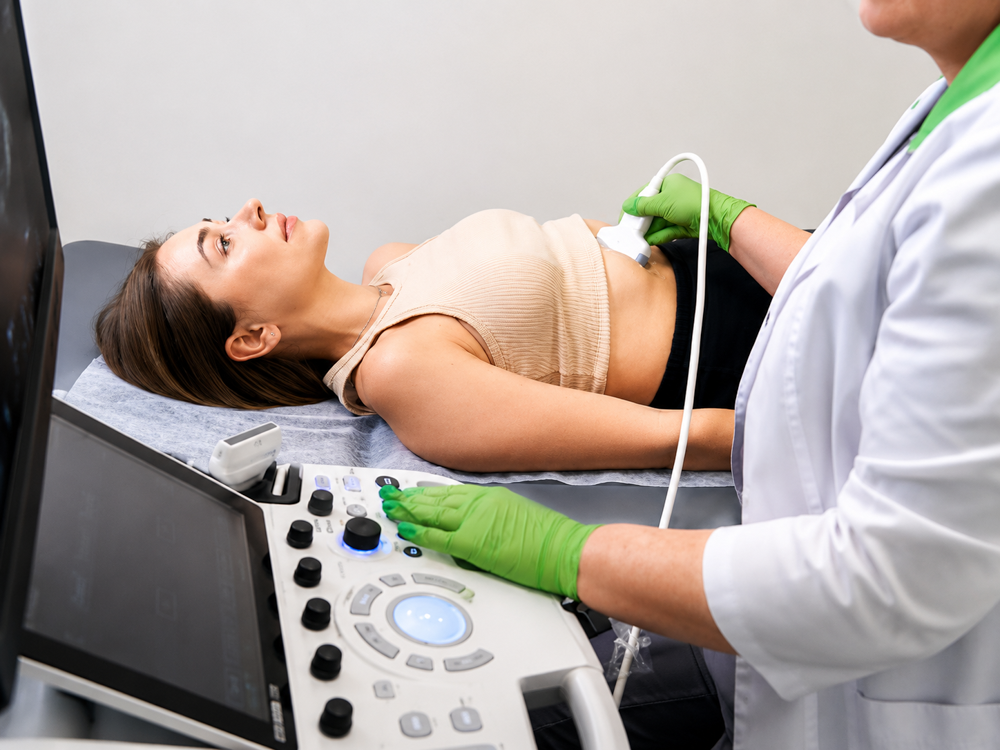
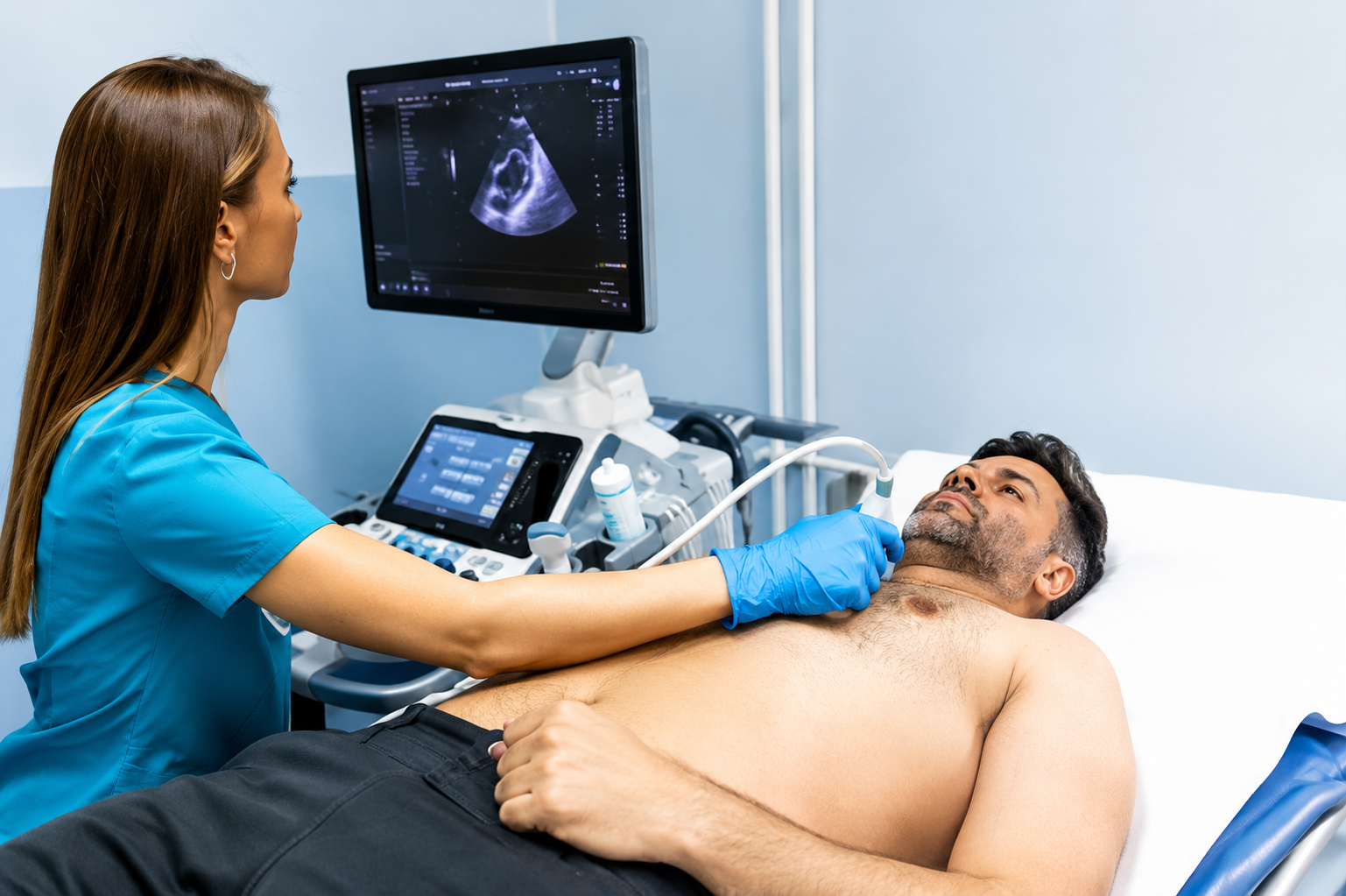
During an echocardiogram, ultrasound images of the heart are obtained while you lie comfortably on an examination couch. A handheld ultrasound probe is used to capture real-time pictures of the heart from different positions on the chest.
The examination generally involves several steps:
- You will be asked to lie in a suitable position.
- Ultrasound gel will be applied to the chest area.
- The ultrasound probe will be placed against the skin.
- Images of the heart will be obtained from different viewing angles.
- The healthcare professional may ask you to change position slightly during the examination.
- Additional measurements or image recordings may be obtained when necessary.
- The captured images are reviewed and documented for reporting.
The ultrasound probe sends sound waves into the body and receives returning echoes, which are then converted into moving images of the heart.
Because the heart is visualised while it is beating, the examination can provide detailed information about cardiac motion and function that cannot be obtained through a physical examination alone.
The patient remains awake throughout the procedure, and the test is generally completed without the need for injections as part of a routine transthoracic echocardiogram.
Is an Echo Test Painful?
An Echo Test is generally considered a non-invasive examination and is not usually painful. The ultrasound images are obtained by placing a probe on the chest surface rather than inserting instruments into the body.
Most patients experience only:
- Mild pressure from the ultrasound probe
- Sensation of cool ultrasound gel
- Minor discomfort from maintaining a particular position for a short period
If a specific area of the chest is already tender because of an underlying condition, you may notice temporary discomfort when pressure is applied with the probe.
If a specific area of the chest is already tender because of an underlying condition, you may notice temporary discomfort when pressure is applied with the probe.
For most patients, the examination is completed comfortably without significant discomfort.
Does an Echocardiogram Use Radiation?
No. An echocardiogram does not use ionizing radiation. The examination creates images using ultrasound waves rather than X-rays.
This is one of the reasons echocardiography differs from imaging tests such as:
- X-rays
- CT scans
- Other examinations that use ionizing radiation
Instead of radiation, the test relies on sound waves that travel through the body and return as echoes, allowing images of the heart to be produced.
Because the technology is different, an echocardiogram provides information about the heart's structure and movement without exposing the patient to ionizing radiation as part of the examination.
Understanding this distinction is important because many patients mistakenly assume that all imaging tests use radiation, which is not the case with routine cardiac ultrasound.
What Can an Echo Test Show About Heart Function?
One of the main purposes of an echocardiogram is to assess how effectively the heart is working as a pump. Because the examination produces moving images, healthcare professionals can observe how the heart contracts, relaxes and circulates blood during each heartbeat.
The test can provide information about:
- How the heart muscle moves
- Overall pumping performance
- The size of the heart chambers
- THow different parts of the heart work together
- Blood flow through the heart
- Functional changes that may affect cardiac performance
Unlike tests that focus mainly on electrical activity, an echocardiogram allows direct visual assessment of how the heart functions mechanically.
This information can be valuable when evaluating symptoms, monitoring known cardiac conditions or assessing changes in heart performance over time.
The findings are interpreted as part of the overall clinical picture and should not be viewed independently from your symptoms or medical history.
Can an Echocardiogram Detect Heart Valve Problems?
Heart valves play an important role in directing blood flow through the heart, and an echocardiogram is one of the key examinations used to assess how these valves are functioning.
During the test, healthcare professionals can observe the movement of the valves as they open and close throughout the cardiac cycle.
The examination may provide information about:
- Valve structure
- Valve movement
- Blood flow across the valves
- Whether a valve appears to be opening normally
- Whether additional assessment of valve function is required
Because the valves work continuously with every heartbeat, changes in valve performance can sometimes affect how efficiently blood moves through the heart.
An echocardiogram allows these structures to be assessed in real time, helping doctors understand how the valves contribute to overall heart function.
Valve-related findings are interpreted together with symptoms, examination findings and other investigations rather than being assessed in isolation.
What Is the Difference Between an ECG and an Echo Test?
Although both tests are commonly used in cardiac assessment, they provide very different types of information about the heart.
An ECG records the heart's electrical activity, while an echocardiogram creates ultrasound images of the heart itself.
| ECG | Echo Cardiogram |
|---|---|
| Records electrical signals | Produces moving heart images |
| Assesses heart rhythm and electrical activity | Assesses heart structure and function |
| Performed using electrodes on the skin | Performed using ultrasound imaging |
| Provides an electrical recording | Provides visual assessment of the heart |
| Does not directly show heart chambers or valves | Can visualise chambers and valves |
Because the two tests answer different clinical questions, a doctor may recommend one or both depending on the patient's symptoms and medical situation.
An ECG and an Echo Test are often complementary rather than interchangeable.
Can an Echo Test Detect Every Heart Problem?
No. While an echocardiogram provides valuable information about heart structure and function, it cannot identify every possible heart condition on its own.
Every cardiac investigation has strengths and limitations. An Echo Test is particularly useful for evaluating the heart's anatomy, movement and pumping function, but some clinical questions may require additional investigations.
Depending on the situation, a doctor may consider:
- ECG findings
- Exercise-based testing
- Additional imaging examinations
- Blood tests
- Clinical assessment
- Other specialised cardiac investigations
For example, some heart conditions involve electrical abnormalities, while others may require different imaging techniques or additional testing to obtain the necessary information.
A normal echocardiogram therefore does not automatically mean that every possible cardiac disorder has been excluded.
The most accurate interpretation comes from combining the Echo findings with symptoms, medical history and other relevant examinations.
How Much Does an Echo Cardiogram Cost in Padur?
The cost of an Echo Cardiogram in Padur can vary depending on the diagnostic centre and the type of echocardiographic examination being performed.
Before scheduling an appointment, it is useful to confirm:
- Current examination charges
- Appointment availability
- Any preparation requirements
- Whether previous reports should be brought
- The exact cardiac test recommended by your doctor
Because pricing can change over time, the most reliable approach is to contact the diagnostic centre directly for the current fee.
For the latest Echo Cardiogram pricing at AVR Scan Centre, contact the Padur centre directly rather than relying on older online information.
What Should You Check Before Booking an Echo Test?
Before arranging an appointment, make sure you understand why the examination has been recommended and which cardiac test your doctor has requested.
It can be helpful to have:
- Your prescription or referral, when applicable
- Previous echocardiogram reports
- Earlier ECG records
- Relevant cardiac investigation reports
- Information about current symptoms
- Details of ongoing cardiac treatment, if relevant
You should also confirm:
- Appointment availability
- Current examination charges
- Any preparation instructions
- Whether previous imaging should be brought for comparison
Bringing relevant earlier reports can sometimes help doctors compare current findings with previous assessments of the heart.
What Happens After an Echo Test?
After the echocardiogram is completed, the recorded images and measurements are reviewed so that the findings can be documented in a formal report. The examination itself may be finished within a relatively short period, but interpretation involves assessing multiple aspects of the heart rather than looking at a single image.
The review process may include:
- Assessment of the heart chambers
- Evaluation of heart muscle movement
- Review of valve appearance and function
- Analysis of blood-flow patterns when applicables
- Measurement of selected cardiac structures
The findings are then organised into a report that helps the referring doctor understand what was observed during the examination.
Depending on why the test was requested, the report may be considered alongside previous cardiac investigations, physical examination findings and current symptoms.
An echocardiogram is most useful when interpreted as part of the complete clinical picture rather than as a standalone result.
An Echo Report is interpreted by evaluating how the heart appears and functions during the examination. The report may contain observations, measurements and an overall impression based on the images obtained.
Depending on the examination, the report may describe:
- Heart chamber characteristics
- Heart muscle movement
- Overall pumping performance
- Valve-related observations
- Blood-flow findings
- Other relevant cardiac features visualised during the test
Some reports contain technical measurements that can be difficult for patients to interpret without clinical context.
For this reason, a report should not be judged solely on the basis of one measurement or medical term. A finding that appears concerning when viewed in isolation may have a different significance when considered alongside the patient's age, symptoms, medical history and other investigations.
Your doctor is best placed to explain what the report means in relation to your individual situation.
What Does an Abnormal Echo Result Mean?
An abnormal Echo result simply means that the examination identified a finding that may require clinical interpretation or further assessment. It does not automatically indicate a serious heart condition.
- Heart structure
- Heart function
- Valve-related observations
- Chamber-related changes
- Other cardiac features visible on ultrasound
Some abnormalities may already be known and are being monitored over time, while others may be newly identified during the examination.
The significance of a finding depends on several factors, including:
- Symptoms
- Medical history
- Physical examination
- Previous cardiac investigations
- Associated risk factors
In some situations, the doctor may recommend follow-up imaging or additional cardiac testing to obtain further information.
An abnormal report should therefore be viewed as part of the diagnostic process rather than as a final diagnosis by itself.
Why Choose AVR Scan Centre for an Echo Cardiogram in Padur?
When searching for an Echo Cardiogram in Padur, patients often look for a diagnostic centre that provides access to cardiac imaging close to home while offering convenient appointment scheduling.
AVR Scan Centre provides Echo Cardiogram services in Padur for patients from Padur, OMR and surrounding areas who require cardiac ultrasound examinations.
The centre also offers a range of diagnostic imaging and cardiac investigations that may be relevant when additional testing is recommended by a healthcare professional.
Before booking, patients can confirm:
- Appointment availability
- Current examination charges
- Preparation requirements
- Reporting-related information
- Any documents or previous reports that should be brought to the appointment
For the most accurate and up-to-date service information, contact AVR Scan Centre directly.
Echo Cardiogram in Padur – Questions Patients Commonly Search
Many routine echocardiograms do not require special dietary restrictions. However, preparation can vary depending on the type of cardiac examination being performed. Follow the instructions provided by your doctor or the testing centre.
The duration can vary depending on the examination and the information that needs to be recorded. Factors such as image quality requirements and the complexity of the assessment can influence the total time.
You can generally communicate with the healthcare professional during the test when necessary. At certain moments, you may be asked to remain still or follow simple instructions so that clear images can be obtained.
Yes, when available. Earlier echocardiograms, ECGs or other cardiac records can sometimes help doctors compare current findings with previous assessments.
Yes, if there is a clinical reason to perform another examination. Follow-up echocardiograms may be recommended when a doctor wants to monitor changes in heart structure or function over time.
Echocardiography can be used in children when clinically appropriate. The specific type of cardiac evaluation depends on the child's condition and the information required by the treating doctor.
Many patients are able to resume their normal routine after the examination. However, follow any specific advice provided by the healthcare professional or referring doctor.
Yes. The term "heart scan" can refer to different types of cardiac imaging examinations. An echocardiogram specifically uses ultrasound to create moving images of the heart.
Book an Echo Cardiogram at AVR Scan Centre, Padur
If your doctor has recommended an Echo Cardiogram in Padur, it is helpful to confirm the examination details before your appointment so that the visit proceeds smoothly.
Before attending, confirm:
- Appointment availability
- Current examination cost
- Any preparation instructions
- Whether previous cardiac reports should be brought
- The exact test recommended by your doctor
Contact AVR Scan Centre – Padur to confirm the Echo Test recommended for you and check the latest appointment and pricing details.
AVR Scan Centre – Padur
Godrej Azure Elsa Plaza, #3, Ground Floor, Padur, Tamil Nadu – 603103.
