

X-Ray in Padur
Diagnostic X-Ray Imaging at AVR Scan Centre
If your doctor has recommended an X-ray, you may be looking for an X-Ray in Padur and want to know what the examination can show, which X-ray you may need, how to prepare and what to expect during the visit.
An X-ray is a diagnostic imaging examination that uses X-rays to create images of structures inside the body. It is commonly used to examine bones, joints and the chest and may also be used for other body areas when clinically appropriate.
AVR Scan Centre provides X-Ray services in Padur for patients looking for diagnostic imaging in and around Padur and the OMR region.
The right X-ray depends on the body part being examined, your symptoms, medical history and the clinical question your doctor wants to answer.
What Is an X-Ray Used For?
An X-ray is used to create images that can help healthcare professionals assess bones, joints, the chest and selected other structures.
X-rays are commonly considered when a doctor needs to evaluate:
- A suspected fracture or bone injury
- Joint or bone pain
- Certain chest-related concerns
- Bone alignment after an injury
- Selected spine or other structural problems
An X-ray provides imaging information, but the image itself is not a diagnosis. Your doctor interprets the findings along with your symptoms, examination and medical history.
Where Can You Get an X-Ray in Padur?
AVR Scan Centre provides X-Ray services at its Padur centre for patients looking for diagnostic imaging in and around Padur.
AVR Scan Centre – Padur
Godrej Azure Elsa Plaza, #3, Ground Floor, Padur, Tamil Nadu – 603103.
The Padur centre is an option for patients searching for an X-Ray Centre in Padur, X-Ray Scan in Padur or an X-Ray near OMR.
Before visiting, confirm:
- The exact X-ray examination required
- Availability
- Whether any preparation is needed
- Appointment requirements
- Any instructions specific to your examination
What Can an X-Ray Show?
What an X-ray can show depends on the body part and the type of examination performed.
Bones and Fractures
X-rays are commonly used to assess suspected fractures and other bone injuries. They can help doctors evaluate bone position, alignment and selected structural changes.
Joints
A joint X-ray can show the bones that form the joint and may help assess certain injuries or alignment changes.
Chest
A chest X-ray can create images of the lungs, heart region and other structures within the chest. It may help evaluate selected chest or lung abnormalities.
Spine
Spine X-rays can provide images of the bony structures of the spine for selected clinical indications.
X-rays are not equally useful for every type of tissue or condition. When more detailed soft-tissue or cross-sectional imaging is needed, your doctor may recommend another examination.
When Does a Doctor Recommend an X-Ray?
A doctor may recommend an X-ray when imaging can help investigate a symptom, injury or suspected structural problem.
Common situations include:
- Recent injury or trauma
- Suspected fracture
- Bone or joint pain
- Swelling or reduced movement
- Certain chest symptoms
- Follow-up of a known finding
The recommendation depends on the clinical situation, not simply on the name of the symptom.
For example, persistent pain may sometimes require a different imaging test depending on the body area and what your doctor is trying to evaluate.
Which X-Ray Do You Need?
The type of X-ray depends on the body area your doctor wants to examine.
Chest X-Ray
A chest X-ray is used to create images of structures within the chest and may be recommended for selected respiratory or chest-related concerns.
Bone and Fracture X-Ray
Bone X-rays are commonly used when a fracture or other bone injury is suspected.
Joint X-Ray
Joint X-rays can help assess the bones and alignment around a joint.
Spine X-Ray
A spine X-ray provides images of the bony structures of the spine for selected clinical needs.
Other X-Ray Examinations
Other body regions may also be examined when an X-ray is appropriate for the clinical question.
Use your doctor's prescription or referral to identify the examination you need rather than selecting an X-ray only from an online search.
Is an X-Ray Safe?
Yes. X-rays are an established diagnostic imaging method, but they use ionizing radiation, so the examination should be performed when it is medically appropriate. The amount of radiation varies with the body part examined, the imaging technique and the number of views required.
For a medically indicated X-ray, the expected diagnostic benefit is considered alongside the radiation exposure.
Before the examination, tell the imaging team if you:
- Are pregnant or may be pregnant
- Have recently undergone other imaging
- Have difficulty standing, sitting or maintaining the required position
- Have any relevant medical concern that may affect the examination
The imaging team can provide instructions and precautions based on the specific X-ray being performed.
Can You Have an X-Ray During Pregnancy?
Tell your doctor and the imaging team if you are pregnant or may be pregnant before having an X-ray.
Whether the examination is appropriate depends on the clinical reason for the scan, the body area being examined and the urgency of the situation.
If an X-ray is medically necessary, your healthcare team can assess the benefits and use appropriate precautions. Do not avoid a medically necessary examination solely because you are concerned about radiation; discuss your situation with your doctor.
Does an X-Ray Use Radiation?
Yes. An X-ray uses ionizing radiation to produce diagnostic images.
The radiation exposure is not the same for every X-ray. It can vary according to:
- Body part being examined
- Number of images required
- Imaging technique
- Patient positioning
This is why X-ray examinations are generally performed when the information they can provide is clinically useful.
If you have recently undergone other imaging, you can mention it to your doctor or the imaging team when relevant.
How Should You Prepare for an X-Ray?
Most routine X-rays require little or no special preparation. However, preparation can vary depending on the body part and the specific examination.
Before your appointment:
- Follow any instructions provided for your particular X-ray.
- Wear comfortable clothing where possible.
- Be prepared to remove jewellery, watches or other metallic objects if they could affect the image.
- Carry your doctor's prescription or referral, if applicable.
- Bring previous relevant imaging reports when they may help with comparison.
Some examinations may have additional instructions, so do not assume that every X-ray requires identical preparation.
What Happens During an X-Ray?
During an X-ray, you are positioned so the required body area can be imaged clearly and with the appropriate view.
The process usually involves:
- Confirming the examination requested by your doctor.
- Positioning you according to the body part being examined.
- Adjusting the X-ray equipment to the required position.
- Asking you to remain still while the image is taken.
- Obtaining additional views if they are needed.
For certain chest X-rays, you may be asked to take a deep breath and hold it briefly while the image is captured.
Good positioning and remaining still help reduce motion-related image problems and support clearer images.
Is an X-Ray Painful?
An X-ray itself is generally not painful because routine X-ray imaging is non-invasive.
The main source of discomfort may be the positioning required to obtain the correct image, particularly if you have an injury, fracture or restricted movement.
You may experience some difficulty if you have:
- A recent fracture
- Joint pain
- Swelling
- Limited mobility
- An injury that makes movement painful
Tell the imaging professional if a required position causes significant discomfort. This is especially important after an injury, when unnecessary movement may be difficult.
What Should You Tell the X-Ray Team Before the Examination?
Provide any information that may affect your safety, positioning or the way the examination is performed.
This may include:
- Pregnancy or possible pregnancy
- Recent injury
- Difficulty standing, sitting or lying in a particular position
- Relevant previous imaging
- Any specific instructions from your doctor
Clear information before the examination helps the imaging team plan the procedure appropriately and reduce avoidable discomfort or delays.
What Can an X-Ray Not Show Clearly?
An X-ray is useful for many bone and chest examinations, but it does not show every type of tissue or condition in the same level of detail.
X-rays may be less useful when the main concern involves certain soft tissues, small or subtle abnormalities, or conditions that require cross-sectional imaging.
Depending on the clinical question, your doctor may recommend another investigation such as:
- CT
- MRI
- Ultrasound
A normal X-ray also does not automatically rule out every possible cause of pain or other symptoms.
The right imaging test depends on what your doctor needs to evaluate.
X-Ray vs CT vs MRI – Which Scan Do You Need?
X-ray, CT and MRI use different imaging methods and are chosen for different clinical purposes.
| X-Ray | CT | MRI |
|---|---|---|
| Uses X-rays | Uses X-rays | Uses magnetic fields and radio waves |
| Produces projection images | Produces cross-sectional images | Produces detailed images in multiple planes |
| Commonly used for bones and selected chest imaging | Useful for detailed cross-sectional assessment | Particularly useful for many soft-tissue examinations |
| Usually a relatively quick examination | Can provide detailed imaging of many body regions | Some examinations take longer |
Which One Is Better?
There is no single imaging test that is best for every condition.
Your doctor may choose X-ray, CT or MRI based on:
- Body area
- Symptoms
- Suspected condition
- Previous findings
- Urgency
- Level of detail needed
Can an X-Ray Confirm a Fracture?
X-rays are commonly used to assess suspected fractures and can often show breaks in bones and changes in bone alignment.
However, not every injury is equally visible on an initial X-ray. If symptoms continue despite a normal or unclear X-ray, your doctor may consider further clinical assessment or another imaging test.
This is why the X-ray should be interpreted together with:
- How the injury happened
- Physical examination
- Pain and swelling
- Ability to move the affected area
- Other relevant findings
A normal X-ray does not always exclude every type of injury.

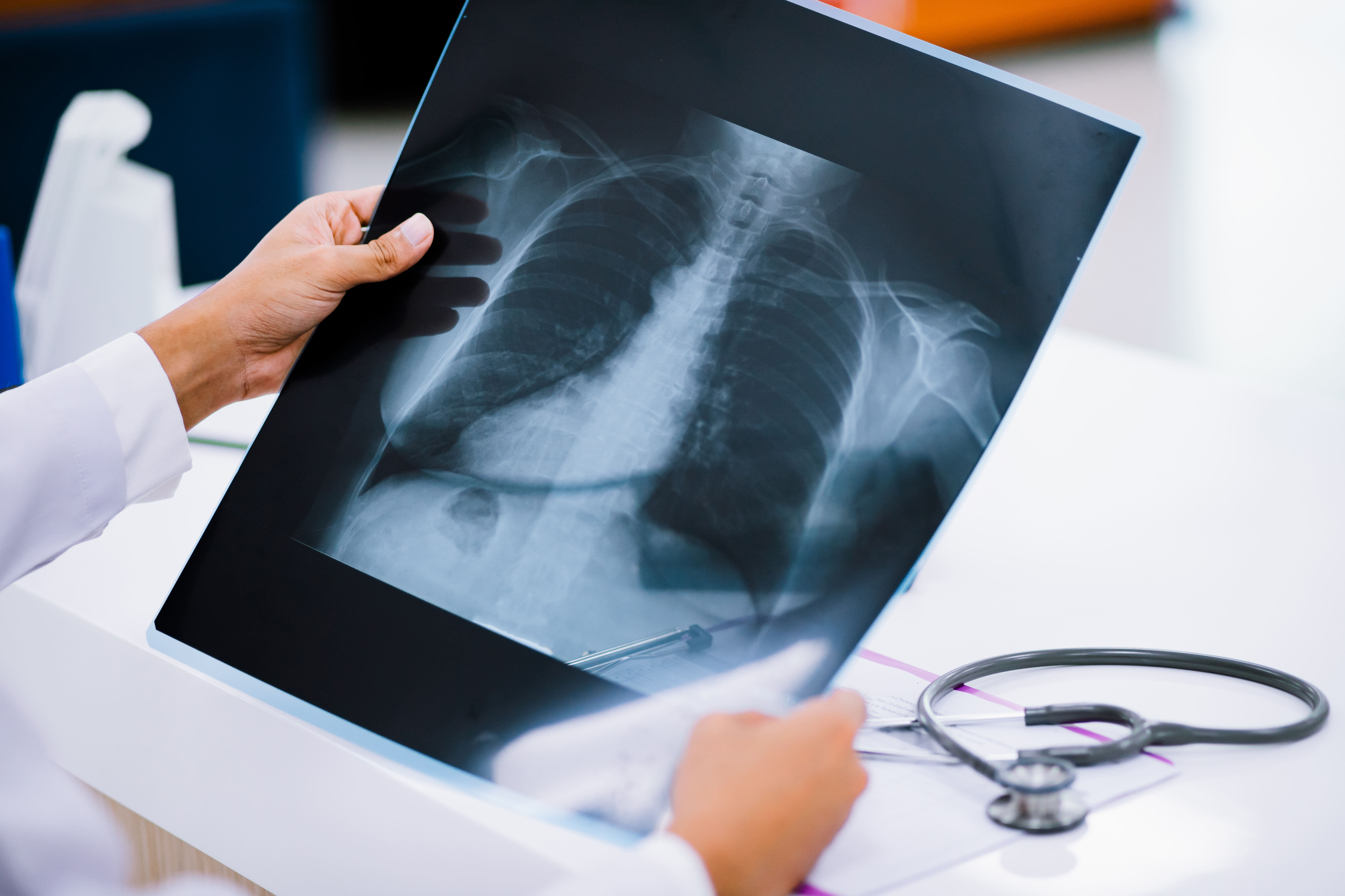
Can a Chest X-Ray Show a Lung Problem?
A chest X-ray can show certain abnormalities in the lungs and chest, but it cannot detect every lung condition.
Depending on the clinical situation, a chest X-ray may help assess selected findings involving:
- Lungs
- Heart region
- Pleural spaces
- Other structures within the chest
Some lung conditions may require additional imaging or testing if the X-ray does not provide enough information.
Your doctor may therefore recommend CT, laboratory testing or another investigation when clinically appropriate.
How Long Does an X-Ray Take?
A routine X-ray is generally a brief examination, although the total appointment time depends on the body part and number of images required.
The visit may take longer when:
- Multiple views are needed
- Positioning is difficult
- The patient has an injury or restricted movement
- Additional images are required for clarification
Ask the centre about the expected appointment time for your specific X-ray.
How Much Does an X-Ray Cost in Padur?
The cost of an X-ray in Padur can vary depending on the body part, type of examination and number of views required.
Before booking, confirm:
- Exact X-ray examination
- Number of views, where applicable
- Current scan charge
- Any additional requirements
For the current price at AVR Scan Centre, contact the Padur centre directly rather than relying on an outdated online price.
What Should You Check Before Booking an X-Ray in Padur?
Before confirming your appointment, make sure you know:
- The exact X-ray examination recommended
- Which body part will be examined
- Whether any preparation is required
- Whether you are pregnant or may be pregnant
- Whether previous imaging should be brought
- Appointment availability
- Current pricing
If your prescription uses a medical term you do not understand, confirm the examination with the centre before booking.
What Happens After an X-Ray?
After the X-ray images are captured, they are reviewed as part of the reporting process.
Your doctor may consider the findings along with:
- Your symptoms
- Medical history
- Physical examination
- Previous imaging
- Other relevant test results
If the X-ray identifies a finding that needs more evaluation, your doctor may recommend another imaging test, laboratory investigation or clinical follow-up.
The X-ray report should be understood in the context of your overall health rather than from an image alone.
What Does an X-Ray Report Tell You?
An X-ray report describes the findings seen on the images and may include observations about the bones, joints, chest or other structures examined.
Depending on the examination, the report may include:
- The body part examined
- Imaging views obtained
- Observed findings
- Measurements, where relevant
- A concluding impression
The meaning of a finding depends on your symptoms and medical history.
If you are unsure about a term or finding in the report, ask your doctor to explain its significance rather than trying to diagnose yourself from the report.
Why Choose AVR Scan Centre for an X-Ray in Padur?
When selecting an X-Ray Centre in Padur, patients may consider the location, availability of the required examination and the process for arranging the scan.
AVR Scan Centre provides X-Ray services in Padur for patients looking for diagnostic imaging in and around Padur and the OMR region.
The Padur centre also provides other diagnostic services that may be relevant when additional investigations are recommended by a doctor.
Any claims about accreditation, radiologist qualifications, specialised equipment, turnaround times, home services or pricing should only be published when verified by AVR Scan Centre.
X-Ray in Padur – Questions People Often Ask
Yes. Previous reports or images can provide useful context and may help with comparison when clinically appropriate.
An X-ray may be performed in children when clinically indicated. The examination should be recommended based on the child's medical need.
Additional X-rays may sometimes be necessary when medically appropriate. Your doctor will determine whether further imaging is needed.
A routine X-ray usually does not require recovery time, so most patients can return to their normal activities unless their underlying condition requires otherwise.
Bring your doctor's prescription or referral, previous relevant imaging and any medical information that may be important for the examination.
Yes. Previous images or reports may help doctors identify changes over time when comparison is clinically useful.
This can depend on the centre's procedures and the type of examination. Check with the centre before your visit if someone needs to accompany you.
Get Your X-Ray at AVR Scan Centre, Padur
If your doctor has recommended an X-ray, knowing the exact examination, preparation requirements and appointment details can help you prepare for the visit.
AVR Scan Centre provides X-Ray services in Padur for patients seeking diagnostic imaging in and around the OMR region.
Before visiting, confirm:
- The required X-ray examination
- Appointment availability
- Preparation instructions, if any
- Current pricing
- Any examination-specific requirements
Need an X-Ray in Padur?
Contact AVR Scan Centre – Padur to enquire about the X-ray examination recommended by your doctor and confirm the requirements for your appointment.
Contact AVR Scan Centre – Padur to enquire about the X-ray examination, preparation requirements and appointment availability.
AVR Scan Centre – Padur
Godrej Azure Elsa Plaza, #3, Ground Floor,
Padur, Tamil Nadu – 603103.
