

Digital Mammogram in Sholinganallur
Digital Breast Imaging at AVR Scan Centre
A Digital Mammogram is an X-ray-based breast imaging examination used to create detailed images of breast tissue.
Digital mammography may be recommended as part of breast screening or when a healthcare professional needs to evaluate a breast-related symptom or finding.
If you are searching for a Digital Mammogram in Sholinganallur, it is useful to understand what mammography is, why it may be recommended, how the examination is performed, how to prepare and how the images are assessed.
A mammogram can help identify certain breast abnormalities that may not be detectable through physical examination alone. However, mammography is not designed to diagnose every breast condition by itself. Additional assessment may sometimes be recommended depending on the findings and clinical circumstances.
What Is a Digital Mammogram?
A Digital Mammogram is a breast X-ray examination that produces digital images of breast tissue for assessment by a qualified radiology professional.
During mammography, the breast is positioned and gently compressed between dedicated imaging surfaces.
Compression helps:
- Spread breast tissue
- Reduce overlapping tissue
- Improve image quality
- Reduce motion during imaging
- Support clearer visualization of breast structures
The examination uses X-rays, so it involves exposure to a small amount of ionizing radiation.
The images are then reviewed as part of the diagnostic or screening process.
Why Is Digital Mammography Performed?
Digital mammography may be performed for breast screening or to evaluate specific breast-related concerns.
A healthcare professional may recommend mammography when there is a need to assess:
- A breast lump or thickening
- Changes in breast appearance
- Changes in the nipple
- Nipple discharge
- Other breast-related symptoms
- A previous breast imaging finding
- Breast health as part of an appropriate screening program
The reason for mammography can differ between individuals.
The decision to undergo mammography should be based on the person's age, symptoms, risk factors, previous findings and healthcare professional's recommendation.
Digital Mammogram for Breast Screening
Mammography is an established breast imaging method used in screening programs to look for certain breast abnormalities in people who may not have noticeable symptoms.
Screening recommendations vary according to factors such as:
- Age
- Personal medical history
- Family history
- Previous breast findings
- Individual risk factors
- Local screening recommendations
There is no single screening schedule that is appropriate for every person.
If you are unsure whether you should have a screening mammogram, discuss your individual circumstances with your healthcare professional.
Diagnostic Mammogram vs Screening Mammogram
The terms screening mammogram and diagnostic mammogram describe different clinical purposes.
Screening Mammogram
A screening mammogram is generally performed when there are no known breast symptoms or specific findings requiring investigation.
Its purpose is to look for breast abnormalities as part of an appropriate screening strategy.
Diagnostic Mammogram
A diagnostic mammogram may be recommended when there is a breast symptom, an abnormal screening result or another specific finding that requires further assessment.
Additional images or other breast imaging may sometimes be recommended depending on the clinical situation.
Your healthcare professional can determine whether screening or diagnostic mammography is appropriate for you.
What Can a Digital Mammogram Show?
A mammogram can show certain changes in breast tissue that may require further evaluation.
Depending on the imaging findings, mammography may help identify:
- Masses
- Calcifications
- Asymmetries
- Architectural changes
- Other breast tissue abnormalities
A finding on a mammogram does not automatically mean that cancer is present.
Some findings may be benign, while others may require additional imaging or further clinical assessment.
The mammogram report should be interpreted by a qualified healthcare professional in the context of the patient's clinical information.

Why Is Breast Compression Necessary During Mammography?
Breast compression is an important part of mammography because it helps spread the breast tissue and reduce movement during image acquisition.
Compression can improve image quality and help reduce the amount of tissue overlap.
It may feel uncomfortable or create pressure for a short period.
If the compression becomes particularly painful, inform the mammography professional.
The examination is designed to keep compression limited to the period necessary to obtain the required images.
How Is a Digital Mammogram Performed?
A digital mammogram involves positioning the breast on the mammography equipment and obtaining X-ray images from different views.
The general process includes:
1. Patient Preparation
The mammography professional confirms the examination and provides instructions.
2. Breast Positioning
The breast is carefully positioned on the imaging platform.
3. Compression
The breast is gently compressed between the imaging surfaces.
4. Image Acquisition
The required mammographic images are obtained.
5. Position Adjustment
The breast may be repositioned to obtain additional views.
6. Image Review
The acquired images are checked for technical quality before the examination is completed.
The exact number of images and views can vary according to whether the examination is for screening or diagnostic evaluation.
Is a Digital Mammogram Painful?
Mammography can cause temporary pressure or discomfort because breast compression is required to obtain the images.
The level of discomfort varies from person to person.
The compression generally lasts only for the time needed to acquire each image.
Tell the mammography professional if you experience significant pain or discomfort during the examination.
How Should You Prepare for a Digital Mammogram?
Most mammograms require relatively simple preparation.
Before the examination:
- Follow the instructions provided by the imaging centre
- Avoid applying deodorant, talcum powder or other products to the breast or underarm area if instructed
- Wear clothing that can be removed easily from the upper body
- Inform the imaging team about previous mammograms
- Bring previous breast imaging reports or images when available
- Inform the healthcare team if you are pregnant or could be pregnant
Previous mammograms can be particularly useful when the radiologist needs to compare current images with earlier examinations.
Can I Have a Mammogram During Pregnancy?
If you are pregnant or think you may be pregnant, inform your healthcare professional before undergoing mammography.
Whether mammography is appropriate during pregnancy depends on the clinical situation and the reason for imaging.
Your healthcare professional will weigh the potential benefits and risks and determine the most appropriate imaging approach.
Do not conceal a possible pregnancy before an X-ray-based examination.
Digital Mammography at AVR Scan Centre
AVR Scan Centre provides Digital Mammography services for patients requiring breast imaging.
If your healthcare professional has recommended a mammogram, confirm the appropriate examination before booking.
Providing previous breast imaging reports or images, when available, can help support comparison with the current examination.
For a Digital Mammogram in Sholinganallur, contact the centre to confirm availability, appointment requirements and preparation instructions.
How Should You Prepare for a Digital Mammogram?
Digital mammography generally requires simple preparation, but following the imaging centre's instructions can help obtain suitable images and avoid unnecessary delays.
Before your appointment:
- Wear clothing that can be removed easily from the upper body.
- Avoid applying deodorant, talcum powder, lotion or other products to the breast or underarm area if instructed by the imaging centre.
- Bring previous mammogram images or reports when available.
- Inform the imaging team about previous breast procedures or breast implants.
- Inform the healthcare professional if you are pregnant or think you may be pregnant.
- Follow any additional instructions provided for your specific examination.
Can I Wear Deodorant Before a Mammogram?
It is generally advisable to avoid deodorant, talcum powder, lotion or similar products on the breast and underarm area before a mammogram if instructed by the imaging centre.
Some products can contain substances that may appear as small areas of increased density or calcification on a mammographic image and potentially complicate image interpretation.
Follow the specific preparation instructions provided when scheduling your examination.
Should I Bring My Previous Mammogram Reports?
Yes. Previous mammograms and breast imaging reports can be useful for comparison with the current examination.
Comparison with earlier images may help the radiologist assess whether a finding is new, unchanged or has changed over time.
If you have previous breast imaging performed at another centre, ask whether you should bring:
- Previous mammogram images
- Previous mammogram reports
- Breast ultrasound reports
- Other relevant breast imaging
- Relevant biopsy or breast procedure records
Having previous information available can support the overall assessment.
How Long Does a Digital Mammogram Take?
The actual image acquisition for a mammogram is usually brief, but the total appointment time can vary.
The duration can depend on:
- Screening or diagnostic purpose
- Number of images required
- Positioning
- Whether additional images are needed
- Patient-specific circumstances
When booking a Digital Mammogram in Sholinganallur, ask the centre about the expected appointment duration.
What Does a Mammogram Feel Like?
Breast compression can cause temporary pressure or discomfort during a mammogram.
The compression is maintained only while the image is being obtained.
The amount of discomfort varies between individuals.
If the examination becomes significantly painful, tell the mammography professional so that the situation can be addressed appropriately.
Is Digital Mammography Safe?
Digital mammography uses X-rays and therefore involves exposure to ionizing radiation.
The examination is performed when the expected diagnostic or screening benefit is considered appropriate.
If you are pregnant or could be pregnant, inform your healthcare professional before the examination so that the imaging decision can be considered in the appropriate clinical context.
As with any medical imaging examination, the suitability of mammography depends on the individual circumstances.
What Happens After a Digital Mammogram?
After the images are obtained, they are assessed by a qualified radiology professional.
The report may describe:
- Breast tissue findings
- Areas of asymmetry
- Masses
- Calcifications
- Other imaging observations
- Comparison with previous examinations, when available
- Recommendations for additional assessment, when appropriate
The exact content of the report depends on the examination and imaging findings.
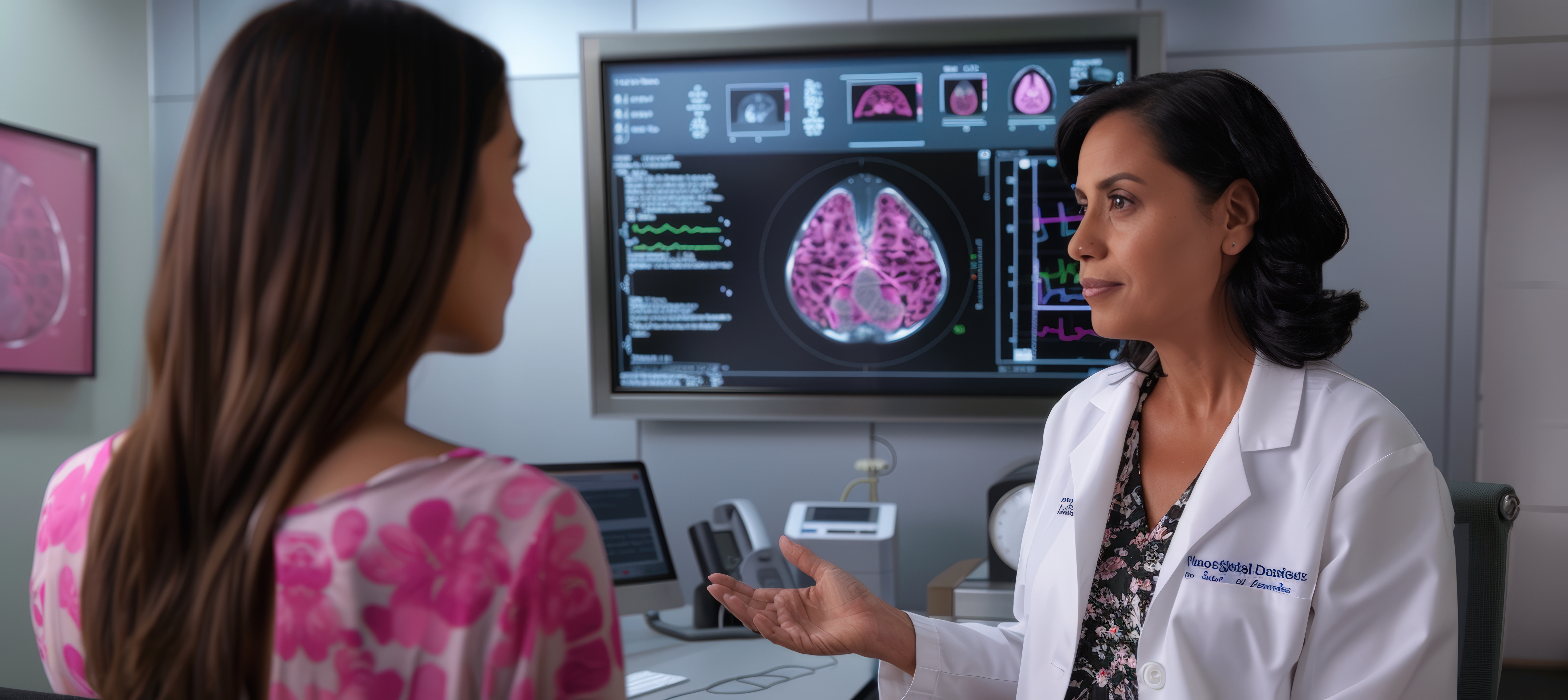
How Is a Digital Mammogram Report Interpreted?
An imaging finding requiring further evaluation does not automatically mean that breast cancer is present. Additional testing may be recommended simply because the mammographic appearance needs further clarification. An abnormal mammogram does not automatically mean that cancer has been diagnosed. A mammogram can show findings that require further assessment, and many findings may ultimately be benign. The next step depends on the specific finding and the overall clinical context. A normal mammogram does not mean that every possible breast condition has been excluded. If you have a persistent lump, nipple discharge, skin changes, pain or another concerning breast symptom, discuss it with your healthcare professional even if your mammogram report is normal. Your doctor may recommend additional clinical assessment or another type of breast imaging depending on the symptom. Do not ignore persistent breast symptoms because of a previous normal imaging report. Mammography and breast ultrasound are different breast imaging examinations and may sometimes be used together. Mammography uses X-rays to create breast images, while ultrasound uses sound waves to evaluate breast tissue. A healthcare professional may recommend additional ultrasound when further assessment is appropriate based on symptoms, mammographic findings or other clinical factors. The choice of imaging depends on the individual's circumstances. If the mammogram identifies an area that requires further evaluation, your healthcare professional may recommend another examination. Additional imaging is not itself a diagnosis. It is a step used to clarify or evaluate a finding. Follow the recommendation provided in your mammogram report or by your healthcare professional. If you are searching for a Mammogram in Sholinganallur, confirm the following before visiting: Providing accurate information about previous breast imaging and relevant medical history can help the imaging team understand the examination requirements. When choosing a Digital Mammogram in Sholinganallur, patients should consider the type of mammography required, preparation instructions, previous imaging availability and how the results will be communicated. AVR Scan Centre provides digital mammography services for patients who have been advised to undergo breast imaging. Digital mammography produces digital X-ray images of the breast that can be reviewed as part of breast screening or diagnostic evaluation. Mammography may be performed for different clinical purposes. Before booking, confirm whether your healthcare professional has recommended a screening mammogram or diagnostic mammogram. If you have had mammography or other breast imaging previously, bringing the relevant images and reports can support comparison with the current examination. The appropriate use of mammography depends on individual circumstances. Do not rely on an online screening schedule alone. Your healthcare professional can determine the most appropriate breast imaging approach based on your individual risk and clinical situation. After receiving your mammogram report, discuss the findings with your healthcare professional, particularly if the report recommends additional assessment. A recommendation for additional imaging does not automatically mean that cancer is present. The purpose of additional assessment is to clarify or investigate a finding. Comparison with previous mammograms can help assess changes in breast tissue over time. For this reason, bring previous mammogram images and reports when available, especially if the earlier examination was performed at another imaging centre. No. Mammography and breast ultrasound are different imaging examinations. Mammography uses X-rays to create images of breast tissue. Breast ultrasound uses sound waves to evaluate breast structures. They can provide different types of information, and a healthcare professional may recommend one or both depending on the patient's age, symptoms, breast tissue characteristics and imaging findings. The appropriate examination should be determined according to the clinical situation rather than choosing an imaging test solely based on an online search. Mammography can help detect certain breast abnormalities that may be associated with breast cancer, but a mammogram alone does not establish every diagnosis. Some findings may require additional imaging or tissue sampling for definitive evaluation. A mammogram may identify features such as masses or calcifications that need further assessment. If your report recommends additional investigation, follow up with your healthcare professional. Breast calcifications are small calcium deposits that can appear on mammographic images. Calcifications can have different appearances and causes. Some may be benign, while particular patterns may require closer assessment. If calcifications are identified, the radiologist may recommend additional mammographic views or other evaluation depending on their appearance. The presence of calcifications does not automatically mean breast cancer. A breast mass identified on mammography may require further evaluation depending on its appearance and clinical context. A mass on a mammogram is not automatically a cancer diagnosis. Your healthcare professional can explain the significance of the finding and recommend the appropriate next step. A Digital Mammogram is an X-ray-based breast imaging examination that produces digital images of breast tissue. It may be used for breast screening or evaluation of specific breast-related concerns. A mammogram can cause temporary pressure or discomfort because the breast needs to be compressed during image acquisition. The compression usually lasts only for the time required to obtain each image. Usually, fasting is not the primary preparation requirement for a mammogram. Follow the specific instructions provided by the imaging centre before your examination. It is advisable to follow the imaging centre's instructions regarding deodorant, talcum powder, lotion and similar products on the breast or underarm area before mammography, as some products can affect image interpretation. Yes. If previous mammogram images or reports are available, bring them or provide access to them when requested. Previous imaging can help with comparison. The image acquisition is generally brief, but the total appointment time varies according to the examination type, number of images and whether additional views are required. Digital mammography uses X-rays and therefore involves exposure to ionizing radiation. Your healthcare professional considers the clinical benefit of the examination when recommending it. Inform the healthcare team if you are pregnant or could be pregnant. An abnormal mammogram does not automatically mean cancer. Additional mammographic views, breast ultrasound, follow-up imaging or biopsy may be recommended depending on the finding. Mammography can be performed in people with breast implants but tell the imaging professional about the implants before the examination. Additional positioning techniques may be required to obtain appropriate breast images. AVR Scan Centre provides Digital Mammography services in Sholinganallur. The centre is located at First Floor, No. 278/1 & 278/2, Commercial Building, Ramanyam Pushkar 2, Kalaignar Karunanidhi Salai, Sholinganallur – 600119. Confirm appointment and examination availability before visiting. If your healthcare professional has recommended mammography, contact AVR Scan Centre to confirm the appropriate examination and appointment availability.
AVR Scan Centre – SholinganallurA report may identify an area that requires:
What Does an Abnormal Mammogram Mean?
Depending on the imaging appearance, your healthcare professional may recommend additional evaluation such as:
What If My Mammogram Is Normal but I Have Breast Symptoms?
Can Mammography and Breast Ultrasound Be Used Together?
What If Additional Breast Imaging Is Recommended?
Depending on the situation, this may include:
Digital Mammogram in Sholinganallur – What Should You Confirm Before Your Appointment?
Why Choose AVR Scan Centre for a Digital Mammogram?
Digital Breast Imaging
Support for Screening and Diagnostic Evaluation
Previous Imaging for Comparison
When Should You Discuss Mammography With a Healthcare Professional?
Discuss breast imaging with your healthcare professional if you have:
What Should You Do After Receiving a Mammogram Report?
Depending on the findings, the next step may involve:
Why Is Comparing Previous Mammograms Important?
A radiologist may compare the current images with earlier examinations to determine whether an imaging finding:
Mammogram vs Breast Ultrasound – Are They the Same?
Can a Mammogram Detect Breast Cancer?
What If a Mammogram Shows Calcifications?
What If a Mammogram Shows a Breast Mass?
Additional assessment may include:
Frequently Asked Questions About Digital Mammogram in Sholinganallur
Book a Digital Mammogram in Sholinganallur
Before your visit, confirm:
For a Digital Mammogram in Sholinganallur, contact the centre to confirm availability, preparation instructions and appointment details.
First Floor, No. 278/1 & 278/2, Commercial Building, Ramanyam Pushkar 2, Kalaignar Karunanidhi Salai, Sholinganallur – 600119.
