
SonoMammography in Sholinganallur
Breast Ultrasound at AVR Scan Centre
If your doctor has recommended SonoMammography, you may want to understand what the examination involves, what it can evaluate, how it differs from other breast imaging tests, whether it is painful, how to prepare and what happens after the examination.
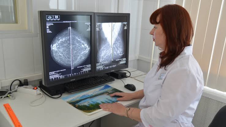
SonoMammography refers to breast imaging that combines mammography and ultrasound assessment of the breast when clinically appropriate. Mammography uses low-dose X-rays to produce images of breast tissue, while breast ultrasound uses sound waves to create images of breast structures.
The two techniques provide different types of information and may complement each other in selected situations.
Breast imaging may be recommended for different reasons, including evaluation of a breast lump, pain, changes noticed in the breast or assessment of an imaging finding.
The appropriate examination depends on factors such as your age, symptoms, clinical history and the reason for the investigation.
If you are looking for SonoMammography in Sholinganallur, AVR Scan Centre provides breast imaging services as part of its diagnostic services.
What Is SonoMammography?
SonoMammography combines mammographic imaging with ultrasound assessment of the breast when both examinations are clinically required.
Mammography
Mammography uses low-dose X-rays to produce detailed images of breast tissue. It can help identify certain breast changes, including abnormalities that may not be easily detected through physical examination alone.
Breast Ultrasound
Breast ultrasound uses high-frequency sound waves to create images of breast tissue. It can provide additional information about a breast finding and may be particularly useful for evaluating whether a finding appears fluid-filled or solid.
Why Are Both Techniques Used?
Mammography and ultrasound provide different information. In some situations, using both techniques can provide a more complete assessment of a breast concern.
However, not every patient requires both examinations. Your doctor or healthcare professional will determine which breast imaging examination is appropriate for your individual situation.
What Can SonoMammography Evaluate?
SonoMammography can be used to evaluate breast findings and provide imaging information about breast tissue.
Depending on the reason for the examination, breast imaging may help evaluate:
- A breast lump
- A palpable breast abnormality
- Breast pain
- Changes in breast tissue
- Certain nipple-related symptoms
- An imaging finding requiring further assessment
- Differences between breast tissue structures
- Other breast concerns identified during clinical evaluation
The purpose of the examination depends on the reason it was requested.
Breast imaging can identify or characterize findings, but an imaging examination alone does not establish the cause of every breast abnormality. If an unusual finding is identified, your doctor may recommend additional assessment.
Why Might a Doctor Recommend SonoMammography?
A healthcare professional may recommend breast imaging when additional information about the breast is needed.
Possible reasons include:
- A breast lump or swelling
- Localized breast discomfort
- A change noticed in the breast
- A finding detected during a clinical examination
- Further evaluation of an imaging finding
- Assessment of breast tissue based on age and clinical circumstances
- Follow-up of a previously identified finding
The appropriate imaging method depends on the individual patient. Your doctor may recommend mammography, breast ultrasound, both examinations or another form of assessment depending on the clinical requirement.
Do not choose a breast imaging examination solely based on an online search.
How Does SonoMammography Work?
SonoMammography involves two different imaging techniques.
How Mammography Works
During mammography, the breast is positioned between specialised imaging plates. A low-dose X-ray is then used to create an image of the breast tissue.
Images are generally obtained from specific views to provide an appropriate assessment of the breast.
Temporary compression is used during mammography to help spread the breast tissue and produce clear images.
How Breast Ultrasound Works
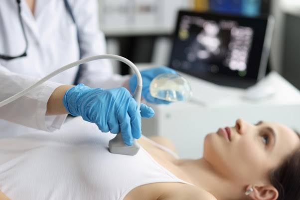
During breast ultrasound, a layer of ultrasound gel is applied to the skin. An ultrasound transducer is moved over the breast area.
The transducer sends sound waves into the breast and receives the returning signals. The ultrasound system processes these signals to create images that can be viewed on a monitor.
What Happens During SonoMammography?
Understanding the examination beforehand can help you know what to expect.
1. Registration and Examination Details
The centre may confirm your details and the type of breast imaging examination requested. You may also be asked about relevant symptoms, previous breast investigations and other information needed for the examination.
2. Mammography
For the mammography portion, the breast is positioned appropriately for imaging. Compression is applied briefly while the images are obtained.
You may experience pressure or temporary discomfort during compression.
3. Ultrasound Assessment
For the ultrasound portion, gel is applied to the skin and the transducer is moved across the breast.
The healthcare professional may examine specific areas based on the clinical indication or findings from the mammographic examination.
4. Image Acquisition
The required images are recorded for assessment and reporting. Additional images may be obtained if needed to evaluate a particular area.
Is SonoMammography Painful?
SonoMammography is generally a non-invasive breast imaging examination, although mammography can cause temporary discomfort because breast compression is required.
The level of discomfort can vary from person to person.
Breast ultrasound itself is generally painless. You may feel mild pressure from the ultrasound transducer as it moves over the breast.
If you experience significant discomfort during mammography or ultrasound, inform the healthcare professional performing the examination.
Does SonoMammography Use Radiation?
The mammography component uses a low dose of X-ray radiation, while ultrasound uses sound waves and does not use ionizing radiation.
Because mammography uses X-rays, inform your healthcare professional if you are pregnant or think you may be pregnant.
The healthcare team can consider this information when determining the appropriate imaging approach.
What Is the Difference Between Mammography and Breast Ultrasound?
Mammography and breast ultrasound use different technologies and provide different types of breast imaging information.
| Mammography | Breast Ultrasound |
|---|---|
| Uses low-dose X-rays | Uses high-frequency sound waves |
| Produces X-ray images of breast tissue | Produces ultrasound images |
| Requires temporary breast compression | Does not require breast compression |
| Can identify certain breast tissue changes and calcifications | Can provide additional information about selected breast findings |
| May be used for screening and diagnostic assessment depending on age and clinical circumstances | Often used as an additional or targeted examination in selected situations |
Neither examination should automatically be considered a replacement for the other. Your healthcare professional will determine which examination or combination of examinations is appropriate.
Can Breast Ultrasound Find a Breast Lump?
Breast ultrasound can provide imaging information about a breast lump or other palpable finding.
It can help characterize a finding and may provide information about whether it appears cystic or solid.
However, ultrasound findings need to be interpreted together with the clinical examination and other relevant breast imaging.
If a breast lump is present, do not rely on an online description to determine whether it is harmless.
Any new, persistent or concerning breast change should be assessed by an appropriate healthcare professional.
Can SonoMammography Detect Breast Cancer?
Breast imaging can identify findings that may require further evaluation, but imaging alone does not confirm every breast cancer diagnosis.
Mammography and ultrasound can identify or characterize certain breast abnormalities.
If an imaging finding is considered suspicious, additional evaluation may be recommended.
Depending on the finding, this may involve:
- Additional breast imaging
- Follow-up imaging
- Clinical assessment
- Further diagnostic testing
- Tissue sampling when clinically indicated
The next step depends on the imaging findings and your healthcare professional's assessment.
A breast imaging report should always be interpreted in the appropriate clinical context.

How Should You Prepare for SonoMammography?
SonoMammography generally requires little preparation, but following the instructions provided for your appointment is important.
Avoid Applying Products to the Breast and Underarm Area
On the day of the examination, you may be advised to avoid applying deodorant, talcum powder, lotion, cream or other cosmetic products around the breast and underarm area.
Some products can affect the quality or appearance of mammographic images. If you are unsure about what to avoid, confirm the instructions with the diagnostic centre before your appointment.
Wear Comfortable Clothing
Wear clothing that can be removed easily from the upper body. You will generally need to remove clothing from the upper body so that the breast imaging examination can be performed appropriately.
Bring Previous Breast Imaging Reports
If you have previously undergone:
- Mammography
- Breast ultrasound
- Breast imaging
- Breast biopsy
- Other relevant breast investigations
Bring the available reports or images if requested. Previous examinations can be useful when your healthcare professional needs to compare current and earlier findings.
Inform the Team About Pregnancy
Tell your doctor and the imaging team if you are pregnant or think you may be pregnant. Mammography uses X-rays, so this information is relevant when determining the appropriate imaging approach.
Tell the Team About Breast Symptoms
Before the examination, mention relevant symptoms such as:
- A new breast lump
- Breast pain
- Nipple discharge
- Skin changes
- A change in breast size or shape
- A recently noticed breast abnormality
Providing accurate information can help the healthcare team understand the reason for the examination.
What Should You Tell the Imaging Team Before a Breast Scan?
Before your SonoMammography in Sholinganallur, provide relevant information about your breast health and previous investigations.
Tell the team if:
- You have noticed a new breast lump
- You have breast pain or tenderness
- You have noticed nipple discharge
- You have had previous breast surgery
- You have breast implants
- You have undergone a previous breast biopsy
- You have had an earlier mammogram or breast ultrasound
- You are pregnant or may be pregnant
- You have been advised to undergo follow-up imaging
This information can help the imaging team understand the examination being performed and the area that may require particular attention.
What Happens During the Mammography Portion?
The mammography component follows a structured imaging process.
Breast Positioning
The breast is positioned carefully on the mammography equipment. The imaging professional will position you appropriately for the required views.
Breast Compression
The breast is briefly compressed between two surfaces. Compression helps spread the breast tissue and can improve the quality of the mammographic image.
It may also help reduce movement during image acquisition.
Image Acquisition
The required mammographic images are obtained. You may be asked to remain still and follow positioning instructions during each image.
Additional Views When Required
Sometimes additional mammographic views may be required to assess a particular area more clearly.
This does not necessarily mean that an abnormality has been found. The additional view may simply be needed to obtain a better image of a specific region.
What Happens During the Breast Ultrasound Portion?
The ultrasound component is performed using an ultrasound transducer.
Application of Ultrasound Gel
A clear ultrasound gel is applied to the skin. The gel helps the transducer move smoothly over the breast and allows ultrasound waves to pass effectively between the transducer and the skin.
Ultrasound Examination
The healthcare professional moves the transducer over the breast and examines the required areas.
If there is a particular lump, area of pain or previously identified finding, the examination may focus on that region.
Imaging of the Underarm Area
When clinically required, ultrasound assessment may also include the axillary or underarm region. The exact area examined depends on the clinical indication.
How Long Does SonoMammography Take?
The duration of a SonoMammography appointment can vary depending on the examinations required and whether additional images are needed.
The mammography and ultrasound portions involve different imaging steps. The total appointment time may therefore depend on:
- Number of mammographic views
- Whether additional views are required
- Whether targeted breast ultrasound is needed
- Number of areas being assessed
- Whether previous findings need further evaluation
For an accurate estimate, contact the centre before your appointment.
What Can You Expect After SonoMammography?
After the imaging examination is completed, the acquired images are reviewed and a report is prepared according to the examination performed.
You can generally resume your usual activities unless your healthcare professional gives you different instructions.
There is usually no recovery period associated with standard mammography or breast ultrasound.
If you experience an unusual or persistent problem after the examination, contact an appropriate healthcare professional.
How Is a SonoMammography Report Used?
A breast imaging report describes the findings identified during the examination.
Depending on the imaging performed, the report may describe:
- Breast tissue appearance
- Areas of asymmetry
- Masses or focal findings
- Cystic or solid-appearing findings
- Calcifications identified on mammography
- Findings requiring comparison with previous examinations
- Other observations relevant to the examination
The report may also indicate whether further assessment or follow-up is recommended.
The wording of a breast imaging report should not be interpreted without considering your symptoms, examination findings and medical history.
If you have questions about your report, discuss it with your doctor.
What If an Abnormality Is Found?
An imaging finding does not automatically mean that breast cancer is present.
Breast imaging can identify many different types of findings. Depending on the appearance of a finding, your doctor may recommend:
- Routine follow-up
- Repeat imaging
- Additional mammographic views
- Targeted ultrasound
- Clinical assessment
- Specialist consultation
- Biopsy or other diagnostic evaluation when necessary
The appropriate next step depends on the specific finding.
Why Is Follow-Up Sometimes Recommended?
Follow-up imaging may be suggested when a finding needs to be monitored over time or when additional information is needed.
A recommendation for follow-up does not by itself establish a diagnosis. Follow the timeframe recommended by your healthcare professional.
When Should You Seek Medical Advice About a Breast Change?
Breast imaging can be useful when a healthcare professional needs to investigate a breast concern.
You should discuss a new or persistent breast change with an appropriate healthcare professional, particularly if you notice:
- A new lump
- Persistent breast pain
- Nipple discharge, particularly if unusual
- A change in nipple appearance
- Skin dimpling or unusual skin changes
- A change in breast size or shape
- A persistent change that is different from your usual breast pattern
Do not wait for an imaging appointment if you have a concerning symptom that requires prompt medical assessment.
SonoMammography for Breast Imaging in Sholinganallur
If your healthcare professional has advised SonoMammography in Sholinganallur, confirming the exact examination before your visit can help you prepare correctly.
AVR Scan Centre provides breast imaging services and also offers other diagnostic investigations that may be recommended depending on the clinical requirement.
Before your appointment, confirm:
- The examination advised
- Whether both mammography and ultrasound are required
- Any preparation instructions
- Whether previous reports should be brought
- Appointment availability
- The expected examination duration
The examination should be selected according to your healthcare professional's recommendation rather than solely on the basis of an online search.
Why Choose AVR Scan Centre for SonoMammography in Sholinganallur?
When choosing a breast imaging centre, patients generally want clear information about the examination available, preparation requirements, location and appointment process.
AVR Scan Centre provides SonoMammography and other diagnostic imaging services in Sholinganallur, allowing patients to enquire about the breast imaging examination recommended for their clinical requirement.
Breast Imaging Services
The centre provides breast imaging services that can include mammography and ultrasound-based assessment, depending on the examination advised. The exact examination should be confirmed before your appointment.
Diagnostic Imaging at One Centre
Depending on the clinical requirement, other diagnostic investigations may also be available at the centre. This can be useful when your healthcare professional has recommended more than one type of diagnostic examination.
Confirm the Examination Before Visiting
Breast imaging requirements can differ between patients. Before visiting, confirm:
- The exact breast imaging examination required
- Whether mammography, ultrasound or both are advised
- Appointment availability
- Preparation requirements
- Whether previous reports or images should be brought
- Any additional instructions for your examination
What Should You Check Before Booking SonoMammography?
Before booking your breast imaging examination, check the following:
- What examination has been recommended
- Whether both mammography and ultrasound are required
- Whether you need to bring previous breast imaging
- Whether you need to bring your prescription or referral
- Whether any preparation is required
- Whether you are pregnant or may be pregnant
- Whether you have breast implants or previous breast surgery
- Whether the examination is available on your preferred date
Bring Previous Breast Imaging When Available
If you have previous mammograms, breast ultrasound reports or other relevant breast imaging, ask whether you should bring them to your appointment.
Earlier imaging may be useful for comparison when assessing a current examination.
Follow Examination-Specific Instructions
Preparation requirements can vary. Do not assume that instructions for one breast imaging examination apply to another.
Follow the instructions provided by the diagnostic centre and your healthcare professional.
Who May Need Breast Imaging?
Breast imaging may be recommended for different clinical reasons. Depending on your age, symptoms, medical history and clinical assessment, your healthcare professional may recommend breast imaging for:
- Assessment of a breast lump
- Evaluation of breast pain
- Investigation of a breast change
- Assessment of a clinical finding
- Further evaluation of an imaging finding
- Follow-up of a previously identified abnormality
- Breast cancer screening when appropriate
The need for breast imaging and the appropriate examination should be determined according to the individual's clinical circumstances.
Frequently Asked Questions About SonoMammography in Sholinganallur
SonoMammography refers to breast imaging involving mammography and ultrasound assessment when both techniques are clinically appropriate. Mammography uses low-dose X-rays, while ultrasound uses sound waves to produce images of breast tissue.
AVR Scan Centre provides breast imaging services at its Sholinganallur centre. Confirm the availability of the specific mammography and ultrasound examination before your appointment.
Mammography involves temporary breast compression, which can cause pressure or short-term discomfort. Breast ultrasound is generally painless, although mild pressure may be felt from the ultrasound transducer.
The mammography component uses low-dose X-rays. Breast ultrasound does not use ionizing radiation because it uses sound waves.
Preparation is generally limited, but follow the instructions given for your examination. You may also be advised to avoid certain products such as deodorant, powder or lotion around the breast and underarm area on the day of mammography.
Breast ultrasound can provide additional imaging information about a breast lump and can help characterize certain findings. The result should be interpreted together with clinical assessment and other relevant imaging.
Mammography and ultrasound can identify breast findings that may require further evaluation. However, imaging alone does not confirm every breast cancer diagnosis. Further assessment may be recommended when a suspicious finding is identified.
Bring your prescription or referral if applicable and previous mammograms, breast ultrasound reports or other relevant breast imaging if available. Also inform the imaging team about previous breast procedures, implants or relevant symptoms.
Tell your doctor and imaging team if you are pregnant or may be pregnant. Mammography uses X-rays, so your healthcare team will determine the most appropriate imaging approach based on the clinical requirement.
The appointment duration varies depending on whether mammography, ultrasound or both are required and whether additional images are needed. Contact the centre for information about the expected duration of your specific examination.
Get SonoMammography at AVR Scan Centre, Sholinganallur
If your healthcare professional has recommended SonoMammography, choosing the correct breast imaging examination and following the appropriate preparation instructions can help make the appointment straightforward.
AVR Scan Centre provides SonoMammography and breast imaging services in Sholinganallur.
Before your visit, confirm:
- Your prescribed breast imaging examination
- Whether mammography and ultrasound are both required
- Appointment availability
- Preparation instructions
- Whether previous reports or images should be brought
- Any additional information required before the examination
SonoMammography in Sholinganallur
AVR Scan Centre – Sholinganallur
First Floor, No. 278/1 & 278/2, Commercial Building, Ramanyam Pushkar 2, Kalaignar Karunanidhi Salai, Sholinganallur – 600119.
Contact the centre before visiting to confirm the availability and appointment requirements for your prescribed breast imaging examination.
