
OPG X-Ray in Sholinganallur
Orthopantomogram at AVR Scan Centre
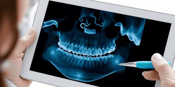
An OPG X-Ray, also known as an Orthopantomogram or panoramic dental X-ray, is an imaging examination that provides a broad view of the teeth, upper and lower jaws and surrounding structures in a single image.
If you are searching for an OPG X-Ray in Sholinganallur, understanding why an OPG may be recommended, what the examination can show, how it is performed and how to prepare can help you know what to expect.
An OPG is commonly used as part of dental and oral assessment. A dentist or other healthcare professional may recommend it when a wider view of the teeth and jaws is needed rather than an image of only one or two teeth.
The appropriate dental imaging examination depends on the clinical question and the area that needs to be assessed.
What Is an OPG X-Ray?
An OPG, or Orthopantomogram, is a panoramic X-ray examination that captures a broad image of the upper and lower jaws and teeth.
Unlike an intraoral dental X-ray, which generally focuses on a smaller area, an OPG provides a wider overview in one examination.
An OPG can include views of:
- Upper teeth
- Lower teeth
- Upper jaw
- Lower jaw
- Jaw joints in the region included by the image
- Surrounding facial structures visible on the panoramic image
The exact structures visible and the quality of assessment depend on positioning, anatomy and the purpose of the examination.
Why Is an OPG X-Ray Performed?
An OPG may be recommended when a dentist or healthcare professional needs a broad radiographic view of the teeth and jaws.
It can be useful when evaluating or planning treatment involving:
- Wisdom teeth
- Tooth development
- Multiple teeth
- Jaw structures
- Dental alignment
- Orthodontic assessment
- Dental treatment planning
- Certain jaw-related findings
The reason for requesting an OPG varies from patient to patient.
An OPG should be performed according to the recommendation of a qualified dental or healthcare professional.
What Can an OPG Show?
An OPG can provide an overview of dental and jaw structures and may help identify findings that require further dental assessment.
Depending on the image quality and clinical situation, an OPG may help assess:
- Number and position of teeth
- Impacted teeth
- Developing permanent teeth
- Wisdom teeth
- General appearance of the jaw bones
- Some dental abnormalities
- Certain areas of bone change
- Structures relevant to orthodontic or dental treatment planning
An OPG does not provide the same level of detail as every other type of dental X-ray.
If a particular tooth or small area requires detailed assessment, a dentist may recommend additional dental imaging.
OPG for Wisdom Teeth Assessment
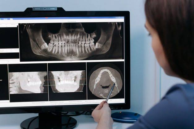
An OPG is commonly used when a dentist needs a broad view of wisdom teeth and their position within the jaws.
It may help show:
- Whether wisdom teeth are present
- Their general position
- Whether they appear impacted
- Their relationship with surrounding teeth
- The overall area of the jaw visible on the panoramic image
If wisdom tooth removal is being considered, the dentist may use the OPG as part of treatment planning.
Additional imaging may sometimes be required when more detailed information is needed.
OPG for Orthodontic Assessment
An OPG may form part of an orthodontic assessment when a broad view of the teeth and jaws is required.
It can provide information about:
- Tooth position
- Missing or unerupted teeth
- Developing teeth
- General dental alignment
- Jaw structures visible on the image
An OPG is only one component of orthodontic assessment. A dentist or orthodontist may require clinical examination and other imaging depending on the treatment plan.
OPG for Dental Treatment Planning
A dentist may recommend an OPG before or during treatment planning when a broad view of the teeth and jaws is useful.
It may be considered for treatment planning related to:
- Wisdom teeth
- Multiple dental problems
- Orthodontic treatment
- Certain oral surgical procedures
- Dental implants, when appropriate
- Jaw-related evaluation
The exact imaging requirements depend on the planned treatment.
An OPG should not be considered a complete assessment of every dental condition.
How Is an OPG X-Ray Performed?
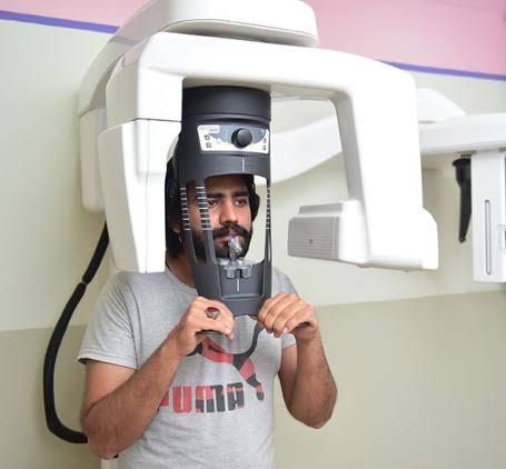
An OPG is performed using dedicated panoramic dental X-ray equipment while the patient remains in a standing or seated position, depending on the equipment.
The general process includes:
1. Preparation
The imaging professional explains the examination and asks you to remove objects that could interfere with the X-ray image.
2. Positioning
You are positioned carefully in the OPG machine. A positioning guide may be used to help keep the head and jaw in the appropriate position.
3. Stabilisation
You may be asked to hold a positioning device or bite on a support so that the teeth and jaws remain correctly aligned during image acquisition.
4. Image Acquisition
The X-ray equipment rotates around the head while acquiring the panoramic image. You will need to remain still during the image acquisition.
5. Image Review
The resulting image is checked for technical quality before the examination is completed.
How Long Does an OPG X-Ray Take?
An OPG X-ray is generally a quick imaging examination, although the total appointment time can vary.
The duration may depend on:
- Patient positioning
- Equipment requirements
- Whether additional images are needed
- Patient movement
- Technical image quality
Following the positioning instructions carefully can help obtain a useful image.
Is an OPG X-Ray Painful?
An OPG is a non-invasive imaging examination and does not normally cause pain.
You may need to bite gently on a positioning device or remain in a particular position for a short period.
If you have difficulty standing, maintaining your position or following the positioning instructions, inform the imaging professional before the examination.
Does an OPG Use Radiation?
Yes. An OPG uses X-rays and therefore involves exposure to ionizing radiation.
The examination should be performed when it is clinically appropriate.
If you are pregnant or think you may be pregnant, inform the healthcare professional before the X-ray examination.
The imaging team can consider the appropriate approach based on the clinical circumstances.
How Should You Prepare for an OPG X-Ray?
An OPG generally requires little preparation.
Before the examination:
- Follow the instructions provided by the imaging centre.
- Remove jewellery or other metallic objects around the head and neck when requested.
- Remove removable dental or facial objects if instructed.
- Inform the imaging professional about relevant dental appliances.
- Inform the healthcare team if you are pregnant or could be pregnant.
- Bring the dentist's prescription or imaging request when applicable.
The imaging professional will provide specific instructions before the examination.
OPG at AVR Scan Centre
AVR Scan Centre provides OPG X-Ray services for patients requiring panoramic dental and jaw imaging.
If your dentist has recommended an OPG, contact the centre before visiting to confirm availability, appointment requirements and any preparation instructions.
For patients searching for an OPG Centre in Sholinganallur, confirming the required examination before booking can help ensure that the appropriate imaging service is arranged.
How Should You Prepare for an OPG X-Ray?
An OPG X-ray generally requires minimal preparation. However, following the imaging centre's instructions is important because metallic objects and incorrect positioning can interfere with the panoramic image.
Before your appointment:
- Remove jewellery around the head and neck when instructed.
- Remove glasses if requested.
- Remove removable dental or facial objects when instructed.
- Inform the imaging professional about dental appliances.
- Tell the healthcare team if you are pregnant or could be pregnant.
- Bring your dentist's prescription or imaging request when applicable.
- Follow any additional instructions provided before the examination.
Why Should Metal Objects Be Removed Before an OPG?
Metallic objects can create artefacts on an X-ray image and may interfere with the assessment of the teeth and jaw structures.
Depending on the equipment and examination, you may be asked to remove:
- Earrings
- Neck chains
- Facial jewellery
- Hair accessories containing metal
- Glasses
- Other removable metallic objects around the head and neck
The imaging professional will tell you which items need to be removed before the scan.
What Happens During an OPG X-Ray?
During an OPG, the patient is positioned carefully in the panoramic X-ray machine while the equipment rotates around the head.
The examination generally follows these steps:
Patient Positioning
You will be positioned in front of the panoramic X-ray equipment. The imaging professional adjusts your head and body position according to the requirements of the examination.
Jaw Stabilisation
You may be asked to bite gently on a positioning support. This helps maintain the correct relationship between the upper and lower teeth during image acquisition.
Remaining Still
You need to remain as still as possible while the equipment rotates around your head. Movement can affect image quality and may make another image necessary.
Image Acquisition
The equipment captures a panoramic X-ray image of the teeth and jaws.
Image Quality Check
The resulting image is checked to determine whether it is technically suitable for the requested examination.
Can Children Have an OPG X-Ray?
Children may undergo an OPG when a dentist or healthcare professional considers panoramic imaging appropriate.
An OPG may be useful in situations involving:
- Developing permanent teeth
- Missing or unerupted teeth
- Wisdom teeth
- Orthodontic assessment
- Certain jaw-related concerns
The decision to perform an X-ray in a child should be based on the clinical need for imaging.
Parents or guardians should inform the imaging team about any relevant medical information and follow the positioning instructions provided.
Can I Have an OPG If I Have Braces?
Patients with braces may be able to undergo an OPG, depending on the purpose of the examination and the type of dental appliance.
Tell the imaging professional about your braces or other dental appliances before the examination.
The imaging team can provide instructions based on the equipment and examination requirements.
Can I Have an OPG With Dental Implants or Crowns?
Dental implants, crowns and other dental restorations can be present when an OPG is performed.
Inform the imaging professional about relevant dental work before the examination.
Metallic dental materials can appear on X-ray images and may affect how particular areas appear.
The dentist or radiologist will interpret the image in the context of the patient's dental history and the purpose of the examination.
Is an OPG Safe During Pregnancy?
If you are pregnant or think you may be pregnant, inform the healthcare professional before undergoing an OPG X-ray.
An OPG uses X-rays and therefore involves ionizing radiation.
The healthcare professional can determine whether the examination is clinically necessary and whether it should be performed based on the individual circumstances.
Do not hesitate to inform the imaging team about a possible pregnancy.
What Happens After an OPG X-Ray?
After the image has been obtained and checked for technical quality, it is reviewed as part of the reporting process.
The resulting image may be assessed for the structures relevant to the clinical question, including the teeth and jaw areas visible on the panoramic image.
The report can then be provided to the referring dentist or healthcare professional.
Your dentist may use the OPG findings together with:
- Dental examination
- Symptoms
- Previous dental records
- Previous imaging
- Treatment history
- Other investigations
How Is an OPG Report Interpreted?
An OPG report should be interpreted together with the patient's dental examination and clinical history.
A panoramic X-ray provides a broad overview, but it does not provide the same level of detail as every other type of dental imaging.
Depending on the findings, the dentist may recommend:
- No additional imaging
- A focused dental X-ray
- Additional panoramic imaging
- Dental examination
- Other diagnostic imaging
- Further clinical evaluation
An OPG finding should not be interpreted in isolation.
What Does an Abnormal OPG Mean?
An abnormal finding on an OPG does not automatically establish a specific diagnosis.
A finding may require further assessment to determine its significance.
For example, the dentist may identify an area involving:
- A tooth
- Wisdom tooth
- Jaw bone
- Tooth development
- Dental alignment
- Other visible structures
Additional imaging or clinical examination may be recommended when the panoramic image does not provide enough information.
Can an OPG Detect Dental Cavities?
An OPG may show some dental changes, but it is not always the preferred imaging examination for detecting every cavity.
Small or early areas of tooth decay may require more focused dental X-rays.
If your dentist suspects decay in a particular tooth, they may recommend an intraoral dental X-ray or another appropriate examination.
The choice of imaging depends on the tooth, symptoms and clinical question.
OPG vs Intraoral Dental X-Ray – What Is the Difference?
An OPG provides a broad panoramic view, while an intraoral dental X-ray generally provides a more focused view of specific teeth or areas.
OPG
Useful when a broad view of the teeth and jaws is required. It can be particularly useful for:
- Wisdom teeth
- Overall dental assessment
- Developing teeth
- Orthodontic planning
- Broad jaw assessment
Intraoral Dental X-Ray
Provides a more focused image of a particular tooth or smaller region. It may be preferred when the dentist needs detailed assessment of a specific area.
The appropriate X-ray depends on what your dentist needs to evaluate.
OPG vs CBCT – Which One Is Better?
OPG and CBCT are different imaging examinations and are used for different clinical purposes.
An OPG provides a two-dimensional panoramic view.
CBCT produces three-dimensional imaging and can provide more detailed spatial information when clinically required.
A dentist may recommend CBCT when three-dimensional information is necessary for a specific treatment or diagnostic question.
CBCT should not automatically replace an OPG. The appropriate examination depends on the clinical requirement.
What Should You Bring for an OPG Appointment?
If your dentist has referred you for an OPG, consider bringing:
- Dentist's prescription or imaging request
- Previous OPG images
- Previous dental X-ray reports
- Relevant dental records
- Information about previous dental procedures
Previous images can sometimes help with comparison or treatment planning.
OPG X-Ray in Sholinganallur – What Should You Confirm Before Visiting?
If you are searching for an OPG X-Ray in Sholinganallur, confirm the following before your appointment:
- OPG availability
- Appointment requirements
- Dentist's prescription or referral requirements
- Preparation instructions
- Whether previous dental images should be brought
- Expected report availability
This can help ensure that the required panoramic dental imaging is available when you arrive.
Why Choose AVR Scan Centre for an OPG X-Ray?
When looking for an OPG X-Ray in Sholinganallur, it is useful to confirm that the imaging service matches the examination requested by your dentist.
An OPG provides a panoramic view of the teeth and jaws and can support dental assessment and treatment planning when a broad radiographic view is required.
Panoramic Dental Imaging
An OPG captures a broad image of the upper and lower teeth and jaw structures in a single examination.
Useful for Dental Treatment Planning
An OPG may be requested for wisdom tooth assessment, orthodontic evaluation, dental treatment planning and other situations where a panoramic view is clinically useful.
Convenient Sholinganallur Location
For patients searching for an OPG Centre in Sholinganallur, AVR Scan Centre provides OPG imaging services at its Sholinganallur centre.
What Should You Do After Receiving an OPG Report?
Your dentist should interpret the OPG findings along with your dental examination and symptoms.
Depending on the report, your dentist may recommend:
- Routine dental follow-up
- A focused dental X-ray
- Additional imaging
- Clinical examination
- Orthodontic assessment
- Oral or maxillofacial evaluation
- Further treatment planning
An OPG report should not be used as a substitute for a dental examination.
When Is Additional Dental Imaging Needed After an OPG?
Additional imaging may be recommended when the panoramic image does not provide enough detail to answer the clinical question.
For example, a dentist may request another examination when detailed assessment of a particular tooth, root or jaw region is required.
Depending on the situation, this could include:
- Intraoral dental X-ray
- Periapical X-ray
- Bitewing X-ray
- Cephalometric imaging
- CBCT
The appropriate examination depends on the area being assessed and the information required by the dentist.
Can an OPG Replace a Regular Dental Examination?
No. An OPG is an imaging examination and does not replace a clinical dental examination.
A dentist may need to evaluate:
- Teeth and gums
- Oral tissues
- Bite and alignment
- Symptoms
- Dental history
- Existing restorations
- Other clinical findings
The OPG is interpreted together with this information.
When Might a Dentist Recommend an OPG?
An OPG may be recommended when a broad view of the teeth and jaws is useful.
Common situations can include:
- Assessment of wisdom teeth
- Orthodontic treatment planning
- Evaluation of unerupted teeth
- Assessment of multiple teeth
- General panoramic dental assessment
- Certain jaw-related concerns
- Selected treatment-planning situations
The exact reason for an OPG varies between patients.
OPG for Wisdom Tooth Removal Planning
An OPG may help the dentist assess the general position of a wisdom tooth and its relationship with nearby structures visible on the panoramic image.
If the dentist requires more detailed information before removal, additional imaging may be recommended.
This is particularly relevant when the clinical question requires more precise three-dimensional information than a panoramic image can provide.
OPG for Orthodontic Treatment
An OPG can provide an overview of tooth position and development that may be useful during orthodontic assessment.
It may help the orthodontist evaluate:
- Missing teeth
- Unerupted teeth
- Developing teeth
- Tooth position
- General jaw structures
An orthodontic assessment may also involve clinical examination and other imaging depending on the treatment plan.
Frequently Asked Questions About OPG X-Ray in Sholinganallur
OPG stands for Orthopantomogram. It is also commonly called a panoramic dental X-ray because it provides a broad image of the teeth and jaws.
An OPG may be performed when a dentist needs a panoramic view of the teeth and jaws. It can be useful for wisdom tooth assessment, orthodontic planning, unerupted teeth and selected dental treatment-planning situations.
No. An OPG is a non-invasive examination and normally does not cause pain. You may need to bite gently on a positioning support and remain still while the image is acquired.
An OPG is generally a quick examination. The total appointment time can vary depending on positioning, equipment requirements and whether additional images are needed.
Usually, fasting is not required for a standard OPG. Follow any specific instructions provided by your imaging centre or dentist.
Yes, an OPG may be performed for patients with braces when clinically required. Tell the imaging professional about your orthodontic appliances before the examination.
Children may have an OPG when a dentist or healthcare professional considers the examination clinically appropriate, such as for assessment of developing or unerupted teeth or orthodontic planning.
An OPG uses X-rays. If you are pregnant or could be pregnant, inform your dentist and imaging professional before the examination so that the clinical need and appropriate approach can be considered.
An OPG can show some dental abnormalities, but it may not provide sufficient detail for every cavity. A dentist may recommend a more focused dental X-ray when detailed assessment of a particular tooth is required.
AVR Scan Centre provides OPG X-ray services in Sholinganallur. The centre is located at First Floor, No. 278/1 & 278/2, Commercial Building, Ramanyam Pushkar 2, Kalaignar Karunanidhi Salai, Sholinganallur – 600119. Confirm availability and appointment requirements before visiting.
Book an OPG X-Ray in Sholinganallur
If your dentist has recommended a panoramic dental X-ray, contact AVR Scan Centre to confirm the OPG service and appointment requirements.
Before visiting, confirm:
- OPG availability
- Appointment requirements
- Dentist's prescription or referral
- Preparation instructions
- Previous dental-image requirements
- Report availability
For an OPG X-Ray in Sholinganallur, contact the centre to confirm availability, preparation requirements and appointment details.
AVR Scan Centre – Sholinganallur
First Floor, No. 278/1 & 278/2, Commercial Building, Ramanyam Pushkar 2, Kalaignar Karunanidhi Salai, Sholinganallur – 600119.
