
Ultrasound Scan in Sholinganallur – Diagnostic Ultrasound at AVR Scan Centre
An ultrasound scan, also called sonography or ultrasonography, is a diagnostic imaging examination that uses high-frequency sound waves to create images of structures inside the body.
Unlike X-ray and CT imaging, ultrasound does not use ionizing radiation. Depending on the type of examination, ultrasound can be used to assess organs, soft tissues, blood flow and developing pregnancy.
If your doctor has recommended an ultrasound scan, the exact type of examination depends on the body area being assessed and the clinical information required.
AVR Scan Centre provides ultrasound imaging services in Sholinganallur for different diagnostic requirements. Before booking, it is useful to confirm the type of ultrasound mentioned on your prescription because preparation and examination methods can vary.
What Is an Ultrasound Scan?
An ultrasound scan uses high-frequency sound waves to produce real-time images of structures inside the body.
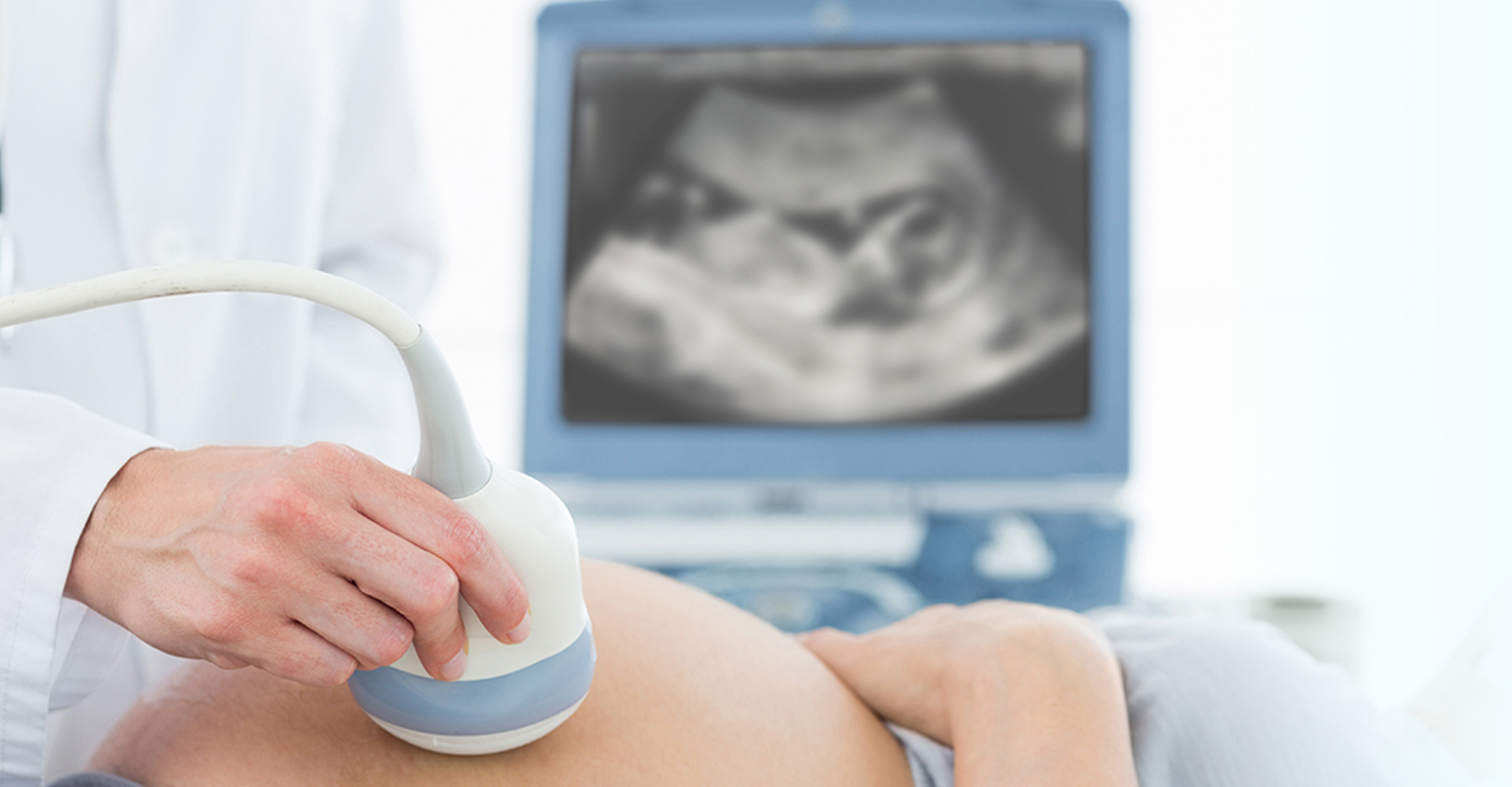
During an ultrasound examination, a device called a transducer sends sound waves into the body. The returning echoes are processed by the ultrasound system to create images.
Depending on the examination, ultrasound can be used to evaluate areas such as:
- Abdomen
- Pelvis
- Urinary system
- Liver
- Gallbladder
- Kidneys
- Pancreas
- Spleen
- Thyroid
- Soft tissues
- Blood vessels
- Developing pregnancy
The area examined and the technique used depend on the doctor's recommendation.
How Does an Ultrasound Scan Work?
Ultrasound imaging works by sending high-frequency sound waves into the body and detecting the echoes that return from different tissues.
The basic process involves:
- Positioning: You are positioned according to the body part being examined.
- Ultrasound gel: A small amount of gel is applied to the skin to help the sound waves travel between the transducer and the body.
- Scanning: The sonographer moves the transducer over the examination area.
- Image creation: Returning sound waves are processed by the ultrasound system into images.
- Image review: The acquired images are documented for interpretation.
Ultrasound can provide real-time imaging, which allows movement and certain dynamic features to be observed during the examination.
What Can an Ultrasound Scan Examine?
The usefulness of ultrasound depends on the body part and clinical question.
Abdominal Ultrasound
An abdominal ultrasound can be used to examine structures within the abdomen, depending on the prescribed examination.
It may include assessment of organs such as the:
- Liver
- Gallbladder
- Kidneys
- Pancreas
- Spleen
The exact organs included depend on the examination requested.
Pelvic Ultrasound
A pelvic ultrasound examines structures within the pelvic region.
Depending on the patient and examination, it may be performed using an abdominal approach or another appropriate technique.
Preparation requirements can differ, so follow the instructions provided for your particular scan.
Kidney and Urinary System Ultrasound
Ultrasound can be used to examine the kidneys and urinary bladder when imaging of the urinary system is clinically required.
It may be recommended as part of an evaluation of urinary or abdominal concerns.
Thyroid Ultrasound
A thyroid ultrasound provides images of the thyroid gland and surrounding soft tissues.
It can help characterize structures within the thyroid when a healthcare professional recommends imaging.
Soft-Tissue Ultrasound
Ultrasound can also be used to examine selected superficial soft tissues.
The area examined depends on the clinical requirement and the doctor's prescription.
Why Might a Doctor Recommend an Ultrasound Scan?
A healthcare professional may recommend ultrasound when imaging of a particular organ, tissue or region can provide useful diagnostic information.
The recommendation may be based on:
- Symptoms
- Medical history
- Physical examination
- Previous investigation results
- The body part requiring assessment
- Pregnancy-related requirements
- The need for real-time imaging
- The specific clinical question
An ultrasound scan should correspond to the examination requested by your healthcare professional.
It is better to follow the prescribed examination rather than choosing an ultrasound type based only on an online search.

Is Ultrasound Safe?
Ultrasound does not use ionizing radiation.
It uses sound waves to create images rather than X-rays.
Ultrasound is widely used for diagnostic imaging, including during pregnancy when clinically appropriate.
However, the examination should still be performed for an appropriate clinical purpose and according to the recommended protocol.
If you are pregnant, have undergone previous imaging or have specific medical concerns, provide the relevant information to the healthcare team before the examination.
What Is the Difference Between Ultrasound and X-Ray?
Ultrasound and X-ray use different technologies and provide different types of information.
| Ultrasound | X-Ray |
|---|---|
| Uses high-frequency sound waves | Uses X-rays |
| Does not use ionizing radiation | Uses ionizing radiation |
| Can provide real-time imaging | Primarily produces projection images |
| Useful for many soft-tissue and fluid-filled structures | Often useful for bones and certain chest examinations |
| Commonly used during pregnancy when clinically appropriate | Pregnancy requires special consideration |
Neither examination is universally suitable for every situation.
The appropriate imaging method depends on the body part, clinical question and recommendation from the healthcare professional.
What Is the Difference Between Ultrasound and CT Scan?
Ultrasound and CT are also different imaging techniques.
Ultrasound uses sound waves and can provide real-time imaging without ionizing radiation.
CT uses X-rays and computer processing to produce detailed cross-sectional images.
Ultrasound may be particularly useful for selected organs, soft tissues, fluid-filled structures and pregnancy-related imaging, while CT can provide detailed cross-sectional imaging of many anatomical regions.
The choice depends on the information required from the examination.
What Types of Ultrasound Examinations May Be Recommended?
The exact ultrasound examination depends on the body area and clinical requirement.
Common categories include:
- Abdominal ultrasound
- Pelvic ultrasound
- Obstetric ultrasound
- Kidney and urinary system ultrasound
- Thyroid ultrasound
- Soft-tissue ultrasound
- Other region-specific ultrasound examinations
Not every examination requires the same preparation.
For example, some abdominal or pelvic examinations may require specific instructions regarding food, fluids or bladder filling.
What Happens During an Ultrasound Scan?
An ultrasound examination is generally a straightforward imaging procedure.
1. Registration and Examination Details
The required examination and relevant patient information are confirmed before scanning.
If you have a prescription, referral or previous relevant report, keep it available.
2. Positioning
You will be asked to lie or sit in a position that allows the required body area to be examined.
The position can vary depending on the type of ultrasound.
3. Application of Gel
A small amount of ultrasound gel is applied to the skin.
The gel helps the transducer make good contact with the skin and allows sound waves to travel effectively.
4. Scanning
The transducer is moved across the area being examined.
You may feel mild pressure as the transducer is moved over the skin, particularly when the examiner needs to obtain images from a specific area.
5. Image Acquisition
Images are captured from different positions or angles as required.
In some examinations, you may be asked to change position, hold your breath briefly or follow another simple instruction.
6. Completion
Once the required images have been obtained, the examination is completed and the images are prepared for reporting.
How Should You Prepare for an Ultrasound Scan?
Ultrasound preparation depends on the type of examination.
There is no single preparation method that applies to every ultrasound scan.
Depending on the examination, you may be asked to:
- Fast for a specified period
- Drink water before the scan
- Have a comfortably full bladder
- Follow dietary instructions
- Take regular medication as advised
- Avoid certain foods before particular examinations
Why Is Preparation Important?
Preparation can help obtain suitable images for certain examinations.
For example, some abdominal examinations may require fasting, while some pelvic or urinary examinations may involve instructions about bladder filling.
Do not assume that fasting or drinking water is required for every ultrasound.
Confirm the preparation instructions for your prescribed examination before visiting the centre.
Is an Ultrasound Scan Painful?
Ultrasound is generally non-invasive and does not usually cause pain.
You may feel pressure when the transducer is moved over the examination area.
The level of pressure can vary depending on the body part being examined and the type of imaging required.
If you experience significant discomfort during the examination, inform the imaging professional.
How Long Does an Ultrasound Scan Take?
The duration of an ultrasound examination varies depending on:
- Type of ultrasound
- Body area being examined
- Number of images required
- Patient positioning
- Whether additional views are needed
- Clinical requirements
Some examinations may be relatively quick, while more detailed examinations can take longer.
For the expected duration of your particular ultrasound, confirm the details when scheduling your appointment.
Can Ultrasound Be Used During Pregnancy?
Yes. Ultrasound is commonly used for pregnancy-related imaging when clinically appropriate.
Pregnancy ultrasound can provide images of the developing pregnancy and may be performed at different stages depending on the purpose of the examination.
The type of obstetric ultrasound recommended depends on factors such as:
- Stage of pregnancy
- Clinical requirement
- Previous findings
- Information the healthcare professional needs
Pregnancy-related ultrasound should follow the examination recommended by your obstetric or healthcare professional.
What Is 3D/4D Ultrasound?
3D ultrasound creates three-dimensional images, while 4D ultrasound adds real-time movement to three-dimensional imaging.
These techniques can provide different ways of viewing anatomical structures during selected examinations.
In obstetric imaging, 3D/4D ultrasound may be recommended when appropriate for the examination.
The availability and purpose of 3D/4D imaging depend on the prescribed examination and clinical requirements.
What Is Doppler Ultrasound?
Doppler ultrasound is an ultrasound technique used to assess blood flow.
It can provide information about the movement of blood through selected blood vessels.
Depending on the examination, Doppler techniques may be used to assess:
- Direction of blood flow
- Relative blood-flow characteristics
- Selected blood vessels
- Circulation in particular anatomical regions
A Doppler examination should be performed when clinically indicated.
Can Ultrasound Detect Every Health Condition?
No. Ultrasound is not suitable for detecting or evaluating every medical condition.
Its usefulness depends on:
- Body part
- Tissue characteristics
- Clinical question
- Patient factors
- Type of ultrasound technique
- Quality of the acoustic window
Certain structures may be difficult to evaluate with ultrasound, and another imaging method may sometimes be recommended.
Your healthcare professional determines which examination is most appropriate for the clinical situation.
What Happens After an Ultrasound Scan?
After the examination, the acquired images are reviewed and documented for reporting.
The report may describe the appearance of the structures examined and any relevant findings identified during the examination.
The findings should be interpreted in relation to:
- Symptoms
- Medical history
- Clinical examination
- Previous investigations
- Other diagnostic reports
An ultrasound report should not be interpreted in isolation.
If you have questions about the findings, discuss the report with the healthcare professional who requested the examination.
Understanding Your Ultrasound Report
An ultrasound report may contain medical terms describing the structures examined and the observations made during imaging.
The report can include information about:
- Size and appearance of organs
- Visible structural features
- Cysts or fluid-filled areas
- Stones where visible
- Measurements
- Blood-flow observations in Doppler examinations
- Pregnancy-related measurements in obstetric examinations
- Other findings relevant to the requested examination
A finding mentioned in an ultrasound report does not automatically establish a diagnosis.
The healthcare professional who requested the examination can interpret the findings alongside your symptoms and medical history.
When Should You Contact the Diagnostic Centre Before Your Scan?
Contact the centre before your appointment if you are unsure about:
- The exact ultrasound examination prescribed
- Whether fasting is required
- Whether you need to drink water
- Whether your bladder needs to be full
- Whether you should continue your regular medication
- Whether previous reports are required
- Whether an appointment is necessary
- The expected duration of the examination
Getting these details beforehand can help prevent unnecessary delays or the need to repeat an examination because preparation requirements were not followed.
Ultrasound Scan in Sholinganallur – What to Know Before Booking
If you are searching for an ultrasound scan in Sholinganallur, the most important step is to identify the exact examination recommended by your healthcare professional.
Different ultrasound examinations have different preparation requirements.
Before booking, confirm:
- The name of the examination
- The body area to be scanned
- Preparation requirements
- Whether Doppler or another ultrasound technique is required
- Whether a pregnancy-related scan has been prescribed
- Appointment availability
- Any documents or previous reports to bring
This helps ensure that the examination performed matches the clinical requirement.
Why Choose AVR Scan Centre for an Ultrasound Scan in Sholinganallur?
AVR Scan Centre provides ultrasound diagnostic imaging services in Sholinganallur for patients who have been advised to undergo ultrasound examinations.
Ultrasound Imaging for Different Diagnostic Needs
Ultrasound can be used for different areas of the body depending on the examination prescribed.
The available service categories can include examinations involving:
- Abdomen
- Pelvis
- Urinary system
- Thyroid
- Soft tissues
- Pregnancy-related imaging
- Other clinically indicated regions
The exact examination should be confirmed before your appointment.
Other Diagnostic Services
The centre also provides diagnostic services including:
- MRI Scan 1.5T
- MultiSlice 3D Spiral CT Scan
- X-Ray
- 3D/4D Obstetrics
- Computerized ECG
- Echocardiogram
- SonoMammography
- Treadmill Test
- EEG
- PFT
- Lab Services
- Preventive Master Health Checkup
- Digital Mammogram
- OPG
This allows patients to enquire about other diagnostic examinations when recommended by their healthcare professional.
Frequently Asked Questions About Ultrasound Scan in Sholinganallur
An ultrasound scan is a diagnostic imaging examination that uses high-frequency sound waves to create images of structures inside the body. It does not use ionizing radiation.
Ultrasound uses sound waves rather than ionizing radiation and is widely used for diagnostic imaging. The examination should be performed when clinically appropriate and according to the recommended protocol.
Ultrasound is generally non-invasive. You may feel some pressure as the transducer is moved over the examination area, but the procedure is generally comfortable.
It depends on the type of ultrasound. Some examinations may require fasting, while others do not. Follow the preparation instructions provided for your specific examination.
Some pelvic and urinary ultrasound examinations may require a full bladder, while others do not. Confirm the preparation requirements for your prescribed scan.
Yes. Ultrasound is commonly used for pregnancy-related imaging when clinically appropriate. The type and timing of the examination depend on the pregnancy and the clinical requirement.
3D ultrasound produces three-dimensional images, while 4D ultrasound adds real-time movement to three-dimensional imaging.
Doppler ultrasound is an ultrasound technique that provides information about blood flow through selected blood vessels or anatomical regions.
The duration depends on the type of examination, body area and images required. Confirm the expected duration when booking your particular ultrasound.
AVR Scan Centre provides ultrasound diagnostic imaging services at its Sholinganallur centre. Contact the centre to confirm the availability of the specific ultrasound examination prescribed by your healthcare professional.
Book an Ultrasound Scan in Sholinganallur
If you have been advised to undergo an ultrasound examination, confirm the exact scan and preparation requirements before your appointment.
Before visiting, confirm:
- Required ultrasound examination
- Appointment availability
- Preparation instructions
- Whether fasting or bladder filling is required
- Whether previous reports should be brought
Contact AVR Scan Centre to confirm your ultrasound appointment and examination requirements.
AVR Scan Centre – Sholinganallur
First Floor, No. 278/1 & 278/2, Commercial Building, Ramanyam Pushkar 2, Kalaignar Karunanidhi Salai, Sholinganallur – 600119.
