
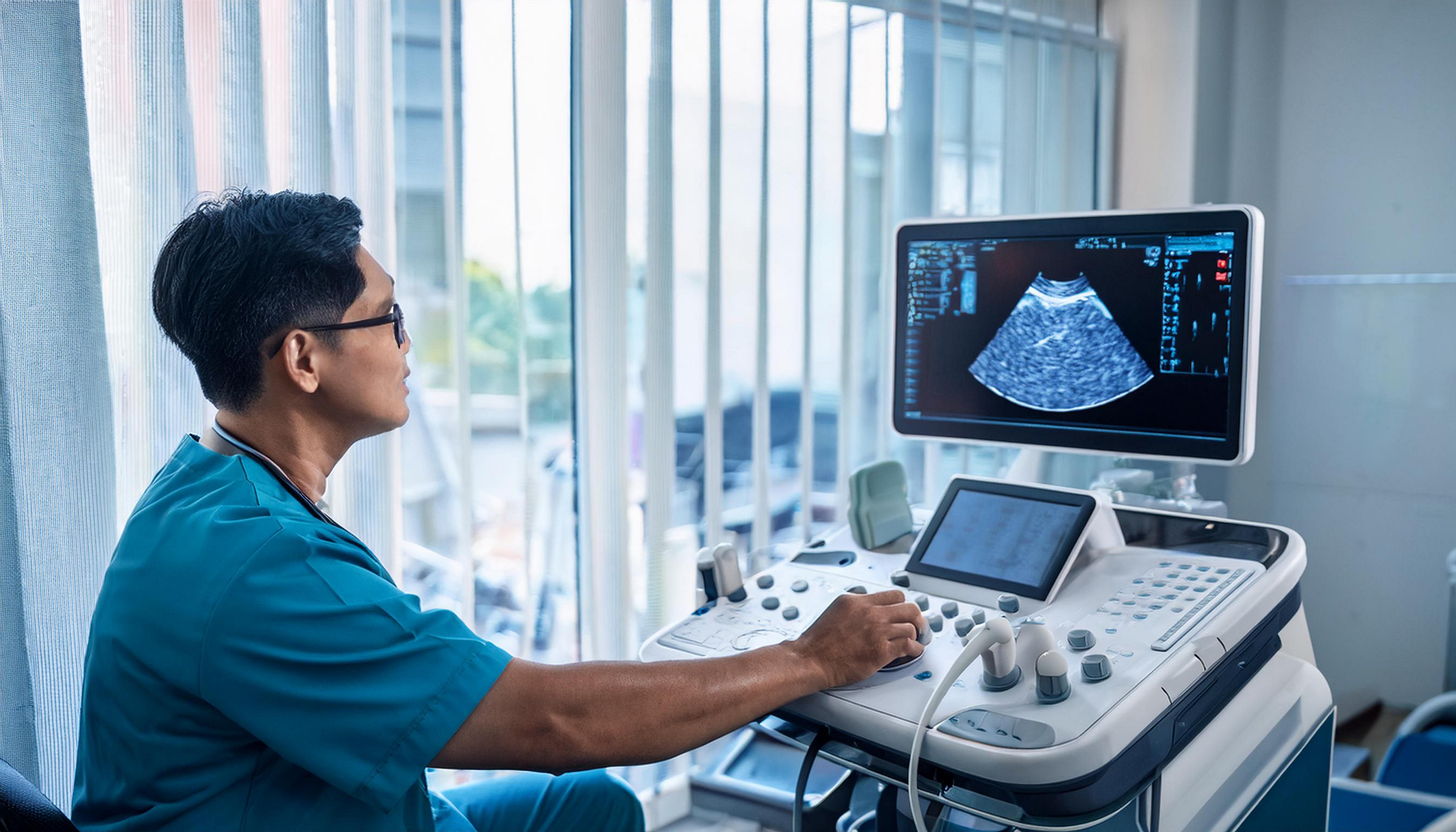
Echo Cardiogram in Sholinganallur
Echo Test at AVR Scan Centre
If your doctor has recommended an Echo Cardiogram, you may want to understand what the test examines, how an echocardiogram is performed, what information it can provide, whether it is painful, how to prepare and what happens after the examination.
An echocardiogram, commonly called an Echo or Echo Cardiogram, is an ultrasound-based examination of the heart. It uses high-frequency sound waves to create moving images of the heart.
Unlike an X-ray-based examination, a standard echocardiogram does not use ionizing radiation.
An echocardiogram can provide information about the heart's chambers, valves, walls and pumping movement. Depending on the type of echocardiographic examination performed, it can also provide information about blood flow through the heart.
If you are looking for an Echo Cardiogram in Sholinganallur, AVR Scan Centre provides echocardiography as part of its diagnostic services.
What Is an Echo Cardiogram?
An Echo Cardiogram is an ultrasound examination that creates images of the heart while it is beating.
The examination uses a device called a transducer, which sends and receives high-frequency sound waves.
The returning sound waves are processed by the ultrasound system to create images of the heart.
An echocardiogram can allow healthcare professionals to assess different aspects of the heart, including:
- Heart chambers
- Heart valves
- Heart walls
- Movement of the heart
- Pumping function
- Blood flow through the heart
- Certain structural features of the heart
The exact information obtained depends on the type of echocardiogram performed and the clinical question being investigated.
What Does an Echocardiogram Check?
An echocardiogram can provide information about the structure and function of the heart.
During an echocardiographic examination, the healthcare professional may assess how the heart chambers and valves appear and how the heart moves during each heartbeat.
Depending on the examination, an echocardiogram may provide information about:
Heart Chambers
The heart contains four chambers that work together to circulate blood. An echocardiogram can provide images of these chambers and may help assess their size and appearance.
Heart Valves
Heart valves help control the direction of blood flow through the heart. Echocardiography can provide images of the valves and can also be used to assess blood flow through them.
Heart Muscle Movement
The movement of the heart walls during contraction can be assessed using echocardiographic imaging.
Pumping Function
An echocardiogram can provide information about how the heart contracts and pumps blood.
Blood Flow
Certain echocardiographic techniques can assess the movement of blood through the heart and across its valves.
Why Is an Echo Cardiogram Done?
A doctor may recommend an echocardiogram when information about the heart's structure or function is required.
The reason for an Echo test varies depending on the patient's symptoms, medical history and clinical requirement.
An echocardiogram may be recommended as part of an evaluation when a patient has symptoms such as:
- Breathlessness
- Chest discomfort
- Palpitations
- Dizziness
- Unexplained tiredness
- Swelling of the legs or feet
- Other symptoms that may require cardiac evaluation
An Echo may also be used when a healthcare professional needs to assess a known or suspected heart condition or monitor changes over time.
The need for an echocardiogram should be determined by your healthcare professional based on your individual clinical situation.
How Does an Echocardiogram Work?
An echocardiogram uses ultrasound waves to create images of the heart.
During a standard echocardiogram, a transducer is placed against the chest.
The transducer sends sound waves into the body and receives the returning echoes.
The ultrasound system processes these signals and displays images of the heart on a monitor.
Because the heart is continuously moving, echocardiography can show the heart in motion rather than producing only a static image.
Depending on the examination, different ultrasound views may be obtained to assess the heart from multiple positions.
What Happens During an Echo Cardiogram?
A standard echocardiogram is generally a straightforward and non-invasive examination.
Preparation for the Examination
You may be asked to remove clothing from the upper body or adjust your clothing to allow access to the chest. The exact preparation depends on the type of echocardiogram being performed.
Positioning
You will generally lie on an examination bed. You may be positioned partly on your side during parts of the examination so that different views of the heart can be obtained.
Electrodes May Be Used
Small ECG electrodes may be placed on the chest during the examination. These can help coordinate the ultrasound images with the heart's cardiac cycle.
Ultrasound Gel Is Applied
A small amount of ultrasound gel is placed on the skin. The gel helps the transducer make good contact with the skin and allows ultrasound waves to travel effectively.
The Transducer Is Moved Across the Chest
The healthcare professional moves the transducer across different areas of the chest to obtain the required views.
You may feel mild pressure as the transducer is positioned.
Images Are Recorded
Different views and measurements may be obtained during the examination. The recorded information is then available for clinical interpretation.
Is an Echo Cardiogram Painful?
A standard echocardiogram is generally painless and non-invasive.
There are no needles involved in a routine transthoracic echocardiogram.
You may feel some pressure when the transducer is pressed against the chest to obtain a clear image.
The ultrasound gel may feel cool on the skin.
If you experience discomfort during the examination, inform the healthcare professional performing the test.
How Should I Prepare for an Echo Cardiogram?
A standard transthoracic echocardiogram usually requires little preparation.
In most cases, you can follow your normal routine unless your healthcare professional has provided specific instructions.
Before your appointment:
- Follow any instructions given by the diagnostic centre.
- Bring your doctor's prescription or referral if applicable.
- Carry previous cardiac reports if they are relevant to your current evaluation.
- Inform the healthcare team about relevant medical information.
- Wear clothing that can be adjusted easily for access to the chest.
If your doctor has requested a particular type of echocardiogram, preparation requirements may differ.
Always follow the instructions provided for your specific examination rather than relying on general preparation advice.
How Long Does an Echo Cardiogram Take?
The duration of an echocardiogram can vary depending on the type of examination and the number of images and measurements required.
A standard echocardiogram may take longer than a basic ECG because multiple ultrasound views of the heart need to be obtained.
The total appointment time can also include preparation, positioning and completion of the examination.
If you need to plan your visit, confirm the expected duration with the diagnostic centre when booking.
Is an Echocardiogram Safe?
A standard echocardiogram uses ultrasound waves and does not use ionizing radiation.
Echocardiography is a non-invasive imaging technique commonly used to examine the heart.
The examination does not involve exposure to X-rays.
The transducer is placed externally against the chest during a standard transthoracic echocardiogram.
If your doctor has recommended an echocardiogram, follow the instructions provided by your healthcare professional and diagnostic centre.
What Is a 2D Echo?
A 2D Echo, or two-dimensional echocardiogram, produces two-dimensional moving images of the heart.
It is a commonly used form of echocardiography.
A 2D echocardiogram can provide views of:
- Heart chambers
- Heart valves
- Heart walls
- Heart movement
- Other visible cardiac structures
Additional echocardiographic techniques may be used when more information about blood flow or cardiac function is required.
The specific type of Echo examination should be based on your doctor's recommendation.
What Is the Difference Between an ECG and an Echo?
An ECG and an echocardiogram are different cardiac investigations.
An ECG records the electrical activity of the heart.
An echocardiogram uses ultrasound to create images of the heart's structures and movement.
| Computerized ECG | Echo Cardiogram |
|---|---|
| Records electrical activity | Produces ultrasound images |
| Provides information about heart rhythm and electrical conduction | Provides information about heart structure and movement |
| Uses electrodes placed on the skin | Uses an ultrasound transducer |
| Does not produce images of the heart's structures | Produces moving images of the heart |
| Usually a short recording | Requires multiple ultrasound views and measurements |
A doctor may recommend an ECG, an echocardiogram or both depending on the clinical information required.
What Information Can an Echo Cardiogram Provide?
An echocardiogram can provide valuable information about the heart's structure and function.
Depending on the examination, it may help assess:
- Size and appearance of heart chambers
- Movement of the heart walls
- Heart valve appearance and movement
- Blood flow through the heart
- Pumping function
- Certain structural abnormalities
- Changes that may require further cardiac evaluation
The findings should always be interpreted in the context of the patient's symptoms and medical history.
An echocardiogram is an investigation and should not be interpreted as a diagnosis without appropriate clinical assessment.
What Happens Before an Echo Cardiogram?
Before the examination begins, the imaging team may confirm your details and the type of echocardiogram requested.
You may be asked about relevant medical information and previous cardiac investigations if they are important to the examination.
For a standard Echo, extensive preparation is usually not required. You will be positioned comfortably so that the required views of the heart can be obtained.
If your examination requires specific preparation, follow the instructions provided when your appointment is scheduled.
What Happens During a 2D Echo?
A standard 2D Echo is performed while you are lying on an examination bed.
The healthcare professional may ask you to turn slightly onto your side during parts of the examination.
Small electrodes may be placed on your chest, and ultrasound gel is applied to the skin.
The ultrasound transducer is then moved across different areas of the chest.
The transducer sends ultrasound waves into the body and receives returning signals. These signals are processed by the ultrasound system to produce moving images of the heart.
Different positions and views may be used to obtain the information required for the examination.
You may be asked to remain still or briefly change your breathing pattern during certain parts of the examination.
What Does an Echo Cardiogram Show?
An echocardiogram provides images that can help assess the heart's structure and function.
Depending on the examination, the images and measurements may provide information about:
- Heart chambers
- Heart valves
- Heart wall movement
- Heart muscle contraction
- Pumping function
- Blood flow through the heart
- Certain structural changes
- Other visible features of cardiac anatomy
The information obtained depends on the type of echocardiogram and the clinical question for which it was requested.
An Echo is therefore different from a test that only records the heart's electrical activity.
Can an Echo Cardiogram Check Heart Valves?
Yes. Echocardiography can provide images of the heart valves and information about blood flow across them.
The heart's valves help direct blood through the chambers of the heart.
During an echocardiographic examination, the appearance and movement of the valves can be assessed.
Doppler techniques may also be used to evaluate blood flow through the heart and across the valves.
If a valve-related abnormality is suspected, your doctor may use the echocardiogram findings together with your symptoms, examination and other relevant investigations.
Can an Echo Cardiogram Check Heart Pumping Function?
An echocardiogram can provide information about how the heart muscle contracts and pumps blood.
The examination produces moving images of the heart, allowing cardiac movement to be assessed.
Measurements may be obtained as part of the examination depending on the type of Echo requested.
If you have been advised to undergo an echocardiogram because of symptoms or a known cardiac condition, the findings should be interpreted together with your overall clinical information.
What Is Doppler Echocardiography?
Doppler echocardiography is an ultrasound technique that can be used to assess the movement of blood through the heart and blood vessels.
While standard echocardiographic imaging shows anatomical structures and movement, Doppler techniques can provide additional information about blood flow.
This can be useful when assessing selected cardiac conditions involving blood flow or heart valves.
The exact Doppler techniques used depend on the type of echocardiographic examination requested.

What Is Color Doppler in an Echo?
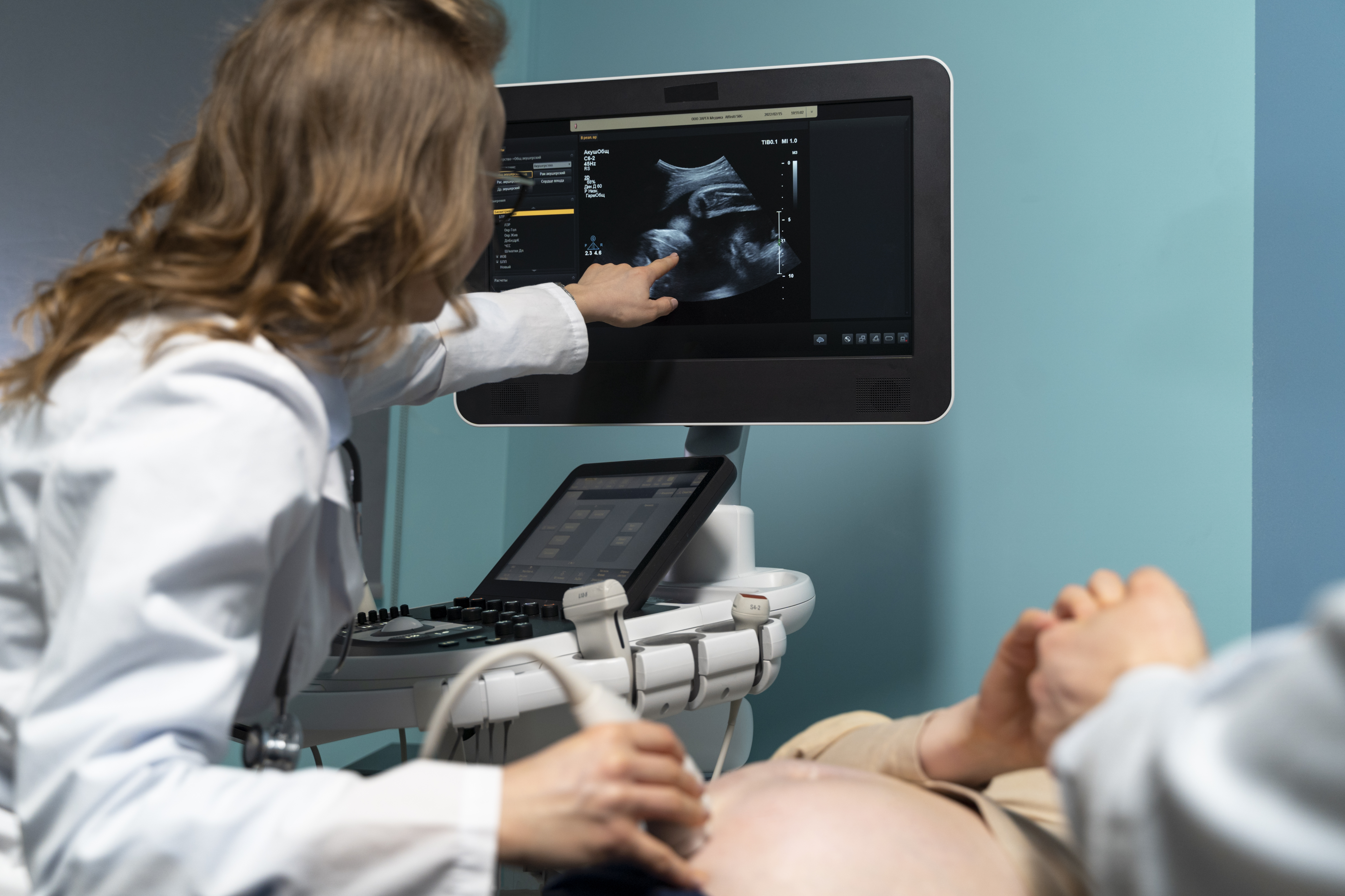
Color Doppler is an echocardiographic technique that displays information about blood flow using colour-coded imaging.
It can help the healthcare professional assess the direction and pattern of blood flow within the heart.
Color Doppler may provide additional information when evaluating selected heart valves and other cardiac structures.
It is not a separate replacement for the basic echocardiographic examination; rather, it can be incorporated into an Echo examination when clinically appropriate.
How Long Does a 2D Echo Take?
The duration of a 2D Echo can vary according to the examination requirements.
The healthcare professional may need to obtain multiple views and measurements of the heart, so the examination can take longer than a standard ECG.
The total appointment time may include:
- Registration
- Preparation
- Positioning
- Electrode placement where required
- Image acquisition
- Measurements
- Completion of the examination
For the expected duration of your particular Echo appointment, contact the diagnostic centre before visiting.
Can I Eat and Drink Before an Echo Cardiogram?
A standard transthoracic echocardiogram generally does not require fasting.
You can usually follow your normal eating and drinking routine unless you have been given different instructions.
However, preparation can vary for different types of cardiac examinations.
If your doctor or diagnostic centre has provided specific instructions, follow those instructions rather than general information available online.
Can I Take My Medicines Before an Echo?
Do not stop prescribed medication simply because you are having an echocardiogram.
If your doctor has provided specific instructions about your medicines before the examination, follow them carefully.
If you are uncertain whether a particular medicine should be taken before your appointment, ask the healthcare professional who prescribed it.
Bring relevant medication information if requested, particularly if the Echo is being performed as part of a wider cardiac assessment.
What Should I Bring for an Echo Cardiogram?
Being prepared can make your appointment easier. If applicable, bring:
- Doctor's prescription or referral
- Previous echocardiogram reports
- Previous ECG reports
- Relevant cardiac investigation reports
- Current medication information
- Other medical records requested by your healthcare professional
Previous reports may be useful when your doctor needs to compare your current findings with earlier investigations.
What Happens After an Echo Cardiogram?
Once the required images and measurements have been obtained, the examination is completed.
For a standard transthoracic Echo, there is generally no recovery period associated with the imaging procedure itself.
You can usually leave after the examination unless you have been given other instructions.
The recorded images and measurements are reviewed as part of the reporting process.
Your doctor can then interpret the findings in relation to:
- Your symptoms
- Medical history
- Physical examination
- Previous cardiac reports
- Other relevant investigations
An echocardiogram report should be considered as part of your overall clinical assessment rather than interpreted in isolation.
What Does an Abnormal Echo Cardiogram Mean?
An abnormal echocardiogram does not automatically mean that a person has a serious heart condition.
The meaning of an Echo finding depends on what was observed, the measurements obtained and the patient's overall clinical circumstances.
Your healthcare professional may consider:
- The specific Echo finding
- Your symptoms
- Medical history
- Previous cardiac investigations
- Current medicines
- Other diagnostic results
Some findings may require further evaluation, monitoring or additional testing, while others may need to be interpreted in the context of the individual patient.
If your Echo report contains an unfamiliar term or abnormal finding, discuss it with your doctor rather than attempting to diagnose yourself from the report.
Can an Echo Cardiogram Detect Every Heart Problem?
No. An echocardiogram does not detect every possible heart condition.
It is primarily an imaging examination that provides information about the heart's structure, movement and, depending on the techniques used, blood flow.
Different heart conditions may require different investigations.
Depending on your symptoms and clinical situation, your doctor may recommend an ECG, Echo, treadmill test, blood tests or other investigations.
The appropriate test should be determined by your healthcare professional based on the information required.
Echo Cardiogram in Sholinganallur – What Should You Know Before Booking?
If you are searching for an Echo Cardiogram in Sholinganallur, it is useful to confirm the exact examination recommended by your doctor before scheduling your appointment.
Before visiting, check:
- Whether a standard 2D Echo has been prescribed
- Whether Doppler or additional echocardiographic assessment is required
- Whether an appointment is necessary
- Whether any preparation instructions apply
- Whether previous cardiac reports should be brought
- Whether you need to carry a prescription or referral
The type of echocardiographic examination should match the clinical requirement rather than being selected solely from an online search.
Echo Cardiogram for Different Cardiac Concerns
An echocardiogram may form part of a cardiac assessment when a healthcare professional needs information about the heart's structure or function.
Depending on the clinical situation, it may be considered when assessing symptoms such as:
- Breathlessness
- Chest discomfort
- Palpitations
- Dizziness
- Unexplained tiredness
- Swelling of the legs
- Other symptoms requiring cardiac evaluation
It may also be used for monitoring when a heart condition has already been identified.
The reason for an Echo test varies from patient to patient, so the examination should be performed according to the healthcare professional's recommendation.
Why Choose AVR Scan Centre for an Echo Cardiogram in Sholinganallur?
When you are looking for an Echo Cardiogram in Sholinganallur, it is useful to choose a diagnostic centre where the required cardiac investigation is available and where you can confirm the examination details before your visit.
AVR Scan Centre provides Echo Cardiogram services in Sholinganallur as part of its diagnostic services.
Echo Cardiogram Service
The centre provides echocardiography for patients who have been advised to undergo an Echo examination.
The test uses ultrasound imaging to provide information about the heart's structure, movement and, where applicable, blood flow.
Other Cardiac Investigations
Depending on the clinical requirement, other cardiac-related investigations available through the centre include:
- Computerized ECG
- Treadmill Test
- Echo Cardiogram
Your doctor may recommend one or more of these investigations depending on the information required.
Diagnostic Services Available
The centre also provides other diagnostic services, including:
- MRI Scan
- MultiSlice 3D CT Scan
- Ultrasound
- X-Ray
- 3D/4D Obstetrics
- EEG
- PFT
- Digital Mammogram
- Laboratory Services
- Preventive Master Health Checkup
The appropriate investigation should always be based on the examination recommended for your individual clinical requirement.
What Should You Check Before Booking an Echo Cardiogram?
Before scheduling an Echo Cardiogram in Sholinganallur, confirm the exact examination your doctor has requested.
It is useful to check:
- Whether a standard 2D Echo has been prescribed
- Whether Doppler assessment is required
- Whether an appointment is necessary
- Whether any preparation is required
- Whether you should bring previous Echo or ECG reports
- Whether you need to bring a doctor's prescription or referral
- The expected duration of the examination
If you have previously undergone an echocardiogram, bringing the earlier report may be useful when your doctor needs to compare findings.
What Can Affect an Echo Cardiogram Examination?
Obtaining clear echocardiographic images can depend on several factors.
The healthcare professional may need to adjust your position or the position of the ultrasound transducer to obtain suitable views.
Factors such as body position, the ability to remain still and the quality of the ultrasound window can influence image acquisition.
If you have difficulty lying in a particular position or experience discomfort during the examination, inform the healthcare professional.
The imaging team can then make appropriate adjustments where possible.
What Happens If More Cardiac Testing Is Needed?
An Echo Cardiogram is one part of cardiac assessment.
Depending on your symptoms and the findings of your examination, your doctor may decide that additional investigations are appropriate.
These may include other cardiac tests such as:
- ECG
- Treadmill Test
- Additional echocardiographic assessment
- Blood investigations
- Other diagnostic examinations
The need for additional testing depends on the individual clinical situation.
A patient should not arrange additional cardiac tests solely because of information found online. Follow the investigation plan recommended by your healthcare professional.
Understanding Your Echo Cardiogram Report
Your echocardiogram report may contain medical terminology, measurements and descriptions of different heart structures.
Some commonly reported areas may include:
- Heart chambers
- Heart valves
- Heart wall movement
- Cardiac function
- Blood flow
- Measurements obtained during the examination
The exact format and terminology of an Echo report can vary according to the examination and reporting system.
If you are unsure about a term in your report, ask your doctor to explain its significance in relation to your symptoms and medical history.
An individual measurement should not be interpreted on its own without the appropriate clinical context.
Why Is an Echo Cardiogram Different From an ECG?
Although both tests are used in cardiac assessment, they provide different types of information.
An ECG records the electrical activity of the heart.
An Echo Cardiogram creates ultrasound images of the heart.
An ECG can provide information about heart rate, rhythm and electrical conduction, while an echocardiogram can provide information about heart structures, movement and blood flow.
Your doctor may recommend either test or both depending on the clinical question.
When Should You Ask About an Echo Cardiogram?
If your doctor has recommended an echocardiogram, you can ask about:
- The reason for the examination
- The type of Echo being performed
- Whether preparation is required
- How long the examination is expected to take
- Whether you should bring previous reports
- When and how the report will be provided
Understanding the examination beforehand can help you arrive prepared and follow the instructions provided by the diagnostic team.
Echo Cardiogram in Sholinganallur
If you are looking for an Echo Cardiogram in Sholinganallur, AVR Scan Centre provides echocardiography as part of its diagnostic services.
First Floor, No. 278/1 & 278/2, Commercial Building, Ramanyam Pushkar 2, Kalaignar Karunanidhi Salai, Sholinganallur, Chennai – 600119.
Before visiting, confirm the required Echo examination, appointment availability and any instructions applicable to your examination.
Frequently Asked Questions About Echo Cardiogram in Sholinganallur
An Echo Cardiogram, or echocardiogram, is an ultrasound examination that produces moving images of the heart. It can provide information about the heart's chambers, valves, walls, movement and, depending on the technique used, blood flow.
AVR Scan Centre provides Echo Cardiogram services in Sholinganallur. The centre is located at First Floor, No. 278/1 & 278/2, Commercial Building, Ramanyam Pushkar 2, Kalaignar Karunanidhi Salai, Sholinganallur, Chennai – 600119. Confirm the availability of the required Echo examination before visiting.
A standard echocardiogram is generally painless and non-invasive. You may feel mild pressure when the ultrasound transducer is placed against the chest to obtain clear images.
No. A standard echocardiogram uses ultrasound waves rather than X-rays, so it does not use ionizing radiation.
A standard transthoracic echocardiogram generally does not require fasting. However, follow any specific preparation instructions provided for your particular examination.
The duration can vary depending on the type of examination, the number of views required and the measurements that need to be obtained. Confirm the expected appointment duration with the centre.
A 2D Echo is a two-dimensional echocardiographic examination that produces moving images of the heart. It can provide information about cardiac structures, movement and function.
Yes. Echocardiography can produce images of the heart valves and can assess blood flow across them using appropriate ultrasound techniques such as Doppler.
Yes. An echocardiogram can provide information about heart muscle contraction and pumping function. The findings should be interpreted together with the patient's clinical information.
If applicable, bring your doctor's prescription or referral, previous Echo or ECG reports and other relevant cardiac investigation reports. Follow any additional instructions provided before your appointment.
Book an Echo Cardiogram in Sholinganallur
If your doctor has recommended an Echo Cardiogram, understanding the examination and preparing according to the instructions provided can make your visit easier.
AVR Scan Centre provides Echo Cardiogram services in Sholinganallur as part of its diagnostic services.
Before visiting, confirm:
- The type of Echo examination required
- Availability
- Appointment requirements
- Preparation instructions
- Documents or previous reports to bring
Looking for an Echo Cardiogram in Sholinganallur?
Contact AVR Scan Centre – Sholinganallur to confirm the required examination and appointment availability.
AVR Scan Centre – Sholinganallur
First Floor, No. 278/1 & 278/2, Commercial Building,
Ramanyam Pushkar 2, Kalaignar Karunanidhi Salai,
Sholinganallur, Chennai – 600119.
