

3D/4D Pregnancy Scan in Thiruporur
Advanced Obstetric Imaging at AVR Scan Centre
Pregnancy is a remarkable journey that involves continuous monitoring of both maternal and fetal health. Ultrasound imaging plays an important role throughout pregnancy by helping healthcare professionals assess fetal development, monitor growth, and evaluate pregnancy progression.
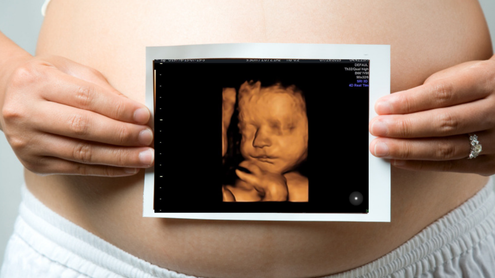
Among the various ultrasound technologies available today, 3D and 4D pregnancy scans provide enhanced visualization of the developing baby compared to conventional two-dimensional imaging. These examinations can offer additional perspectives of fetal anatomy and movement when clinically appropriate.
A 3D/4D pregnancy scan is not a replacement for routine antenatal care. Instead, it may complement standard obstetric ultrasound examinations by providing additional imaging information based on the recommendations of the obstetrician or healthcare provider.
At AVR Scan Centre, Thiruporur, 3D/4D obstetric ultrasound services are available for pregnant women seeking advanced pregnancy imaging as part of their prenatal care journey.
Patients from Thiruporur, Kelambakkam, Padur, Navalur, Siruseri, Chengalpattu, and nearby OMR areas can conveniently access pregnancy ultrasound services.
What Is a 3D/4D Pregnancy Scan?
A 3D/4D pregnancy scan is an advanced form of obstetric ultrasound used to visualize the developing fetus during pregnancy.
Traditional ultrasound creates two-dimensional images that allow healthcare professionals to evaluate fetal anatomy and development.
Advanced imaging technologies can provide additional visualization:
3D Ultrasound
3D ultrasound combines multiple ultrasound images to generate a three-dimensional representation of the baby and selected fetal structures.
This can provide greater surface detail compared to standard 2D imaging.
Advanced imaging technologies can provide additional visualization:
4D ultrasound adds the dimension of real-time movement to 3D imaging.
Instead of viewing a still three-dimensional image, parents and healthcare professionals can observe fetal movements as they occur during the examination.
These technologies may be used when clinically appropriate and when recommended by the healthcare provider managing the pregnancy.
Why Are Pregnancy Ultrasound Scans Important?
Ultrasound examinations play an important role throughout pregnancy because they provide valuable information about fetal growth, development, and overall pregnancy progression.
Doctors may recommend ultrasound examinations to:
- Confirm pregnancy
- Estimate gestational age
- Assess fetal growth
- Evaluate fetal development
- Monitor multiple pregnancies
- Assess placental location
- Evaluate amniotic fluid levels
- Monitor fetal well-being
- Investigate pregnancy-related symptoms
Ultrasound findings are always interpreted together with clinical assessment and other aspects of prenatal care.
What Is the Difference Between 2D, 3D and 4D Pregnancy Scans?
Many parents wonder whether 3D and 4D scans are different from routine pregnancy ultrasounds.
Although all three techniques use ultrasound technology, they provide images in different ways.
| 2D Ultrasound | 3D Ultrasound | 4D Ultrasound |
|---|---|---|
| Flat two-dimensional images | Three-dimensional images | Real-time moving 3D images |
| Standard pregnancy imaging | Enhanced visualization of fetal structures | Live visualization of fetal movement |
| Most commonly used during pregnancy | May be used in selected situations | Provides dynamic fetal imaging |
| Used for routine prenatal assessment | Additional imaging when appropriate | Additional visualization when indicated |
Each technique has a specific role within obstetric imaging.
The type of examination performed depends on the clinical requirement and the recommendation of the healthcare professional.
When Might a Doctor Recommend a 3D/4D Pregnancy Scan?
The timing and need for a 3D/4D ultrasound examination depend on the stage of pregnancy and the clinical situation.
Healthcare providers may recommend advanced obstetric imaging when:
- Additional fetal visualization is useful
- Fetal anatomy requires further assessment
- Follow-up imaging has been requested
- Specific pregnancy-related questions need evaluation
- Enhanced visualization of selected structures is beneficial
The decision regarding imaging should always be guided by the obstetrician or healthcare provider managing the pregnancy.
What Can a 3D/4D Pregnancy Scan Show?
Advanced obstetric ultrasound imaging can provide detailed views of fetal structures and movement.
Depending on fetal position, gestational age, and imaging conditions, the examination may show:
Facial Features
3D imaging may provide visualization of facial contours and surface anatomy.
Fetal Limbs
Arms, legs, hands, and feet may be visualized during the examination.
Fetal Movement
4D imaging can display movement in real time.
Fetal Position
The position of the baby within the uterus can be assessed.
Selected Anatomical Structures
The examination may contribute additional information when evaluating specific fetal structures.
The quality of images obtained depends on several factors, including fetal position, maternal anatomy, gestational age, and amniotic fluid volume.
At What Stage of Pregnancy Is a 3D/4D Scan Usually Performed?
The most appropriate timing for a 3D/4D pregnancy scan varies depending on the purpose of the examination.
Different stages of pregnancy provide different imaging opportunities because fetal size and development change throughout gestation.
Healthcare providers determine the appropriate timing based on:
- Gestational age
- Clinical indication
- Fetal development stage
- Pregnancy history
- Imaging requirements
The obstetrician managing the pregnancy is best placed to advise when advanced ultrasound imaging may be beneficial.
How Does a 3D/4D Pregnancy Scan Work?
3D and 4D ultrasound examinations use the same fundamental ultrasound technology employed in routine obstetric imaging.
A transducer placed on the abdomen sends high-frequency sound waves into the body.
These sound waves reflect from fetal structures and return to the transducer.
Advanced software processes the returning signals to create:
- Three-dimensional images
- Surface-rendered fetal views
- Real-time moving images in 4D mode
The resulting images provide enhanced visualization while maintaining the non-invasive nature of ultrasound imaging.
What Happens During a 3D/4D Pregnancy Scan?
The examination process is similar to a standard obstetric ultrasound.
Registration and Review
Relevant pregnancy information and referral details are reviewed.
Positioning
The expectant mother is positioned comfortably for the examination.
Application of Ultrasound Gel
A water-based gel is applied to the abdomen to improve sound-wave transmission.
Image Acquisition
The ultrasound transducer is moved across the abdomen while images are obtained.
Advanced Imaging
When appropriate, 3D and 4D imaging modes are used to generate enhanced views of the developing baby.
Completion of Examination
Once the required images have been obtained, the examination is completed.
Is a 3D/4D Pregnancy Scan Safe?
One of the most common questions expectant parents ask is whether a 3D/4D pregnancy scan is safe for both mother and baby.
A 3D/4D scan uses the same ultrasound technology that has been widely used in obstetric care for many years. The examination works by transmitting high-frequency sound waves into the body and converting the returning echoes into images.
Unlike X-rays and CT scans, ultrasound imaging does not use ionizing radiation.
Healthcare professionals use obstetric ultrasound when there is a valid medical reason and when the examination contributes to prenatal assessment and pregnancy monitoring.
The scan should always be performed according to established medical guidelines and as recommended by the healthcare provider managing the pregnancy.
Does a 3D/4D Pregnancy Scan Use Radiation?
No.
A 3D/4D pregnancy scan does not use radiation.
Many expectant mothers are concerned about radiation exposure during pregnancy because some medical imaging examinations rely on ionizing radiation.
Ultrasound imaging is different.
Instead of radiation, ultrasound uses sound waves to generate images of the developing baby and surrounding structures.
This is one of the reasons ultrasound remains one of the most commonly used imaging methods throughout pregnancy.
Because there is no radiation exposure, ultrasound plays an important role in routine prenatal monitoring and fetal assessment.
How Should You Prepare for a 3D/4D Pregnancy Scan?
Preparation requirements can vary depending on the stage of pregnancy and the type of examination requested.
In many cases, minimal preparation is required.
However, patients should always follow the instructions provided by the imaging centre or healthcare provider.
Helpful preparation may include:
Stay Hydrated
Adequate hydration may help support ultrasound image quality during certain stages of pregnancy.
Wear Comfortable Clothing
Loose-fitting clothing can make the examination more comfortable.
Bring Previous Pregnancy Records
Previous ultrasound reports and antenatal records can be helpful when comparison is needed.
Arrive on Time
Arriving slightly early allows sufficient time for registration and review of relevant pregnancy information.
If any special preparation is required, patients will usually be informed before the appointment.
Can a 3D/4D Pregnancy Scan Show the Baby's Face?
One of the reasons many parents are interested in 3D imaging is the possibility of seeing detailed facial views of their developing baby.
When imaging conditions are favorable, 3D ultrasound may provide visualization of:
- Facial contours
- Nose
- Lips
- Eyes
- Forehead
- Chin
However, image quality can vary significantly.
Factors such as fetal position, gestational age, amniotic fluid levels, placental location, and maternal body habitus may influence the ability to obtain clear facial images.
For this reason, image appearance varies from one pregnancy to another.
Can a 3D/4D Scan Show Baby Movements?
Yes.
One of the distinguishing features of 4D ultrasound is its ability to display movement in real time.
Depending on fetal activity during the examination, parents may observe:
- Hand movements
- Leg movements
- Stretching
- Facial expressions
- Head movements
- General fetal activity
Because the images are displayed live, 4D ultrasound can provide a dynamic view of fetal movement during the scan.
The amount of movement seen depends entirely on the baby's activity during the examination.
Can a 3D/4D Pregnancy Scan Be Used in Twin Pregnancies?
Twin pregnancies often require more frequent monitoring than singleton pregnancies.
Ultrasound plays a vital role in evaluating fetal growth, development, and pregnancy progression when multiple babies are present.
Depending on fetal position and imaging conditions, 3D/4D ultrasound may be performed in twin pregnancies.
However, obtaining detailed images can sometimes be more challenging because:
- Space within the uterus is shared by multiple fetuses
- Fetal positions may overlap
- One baby may partially obscure the other
- Visualization may vary between examinations
The healthcare provider will determine the most appropriate imaging approach based on the specific pregnancy.
Can a 3D/4D Pregnancy Scan Detect Birth Defects?
Many parents wonder whether a 3D/4D scan can identify fetal abnormalities.
Ultrasound plays an important role in evaluating fetal anatomy during pregnancy.
In selected situations, 3D imaging may provide additional visualization of certain anatomical structures.
However, it is important to understand that:
- No ultrasound examination can identify every abnormality
- Not all conditions are visible on imaging
- Detection depends on gestational age
- Image quality influences assessment
- Some abnormalities may only become apparent later in pregnancy
The purpose of prenatal imaging is to support clinical assessment and fetal evaluation as part of comprehensive obstetric care.
The findings should always be interpreted by qualified healthcare professionals.
What Factors Affect 3D/4D Ultrasound Image Quality?
Many factors influence the quality of images obtained during a pregnancy scan.
Some of the most important include:
Fetal Position
The baby's position inside the uterus can significantly affect visualization.
Gestational Age
Different stages of pregnancy offer different imaging opportunities.
Amniotic Fluid Volume
Adequate fluid around the baby may improve image clarity.
Placental Location
The position of the placenta can sometimes influence imaging.
Maternal Anatomy
Individual anatomical differences can affect ultrasound visualization.
Fetal Movement
Active fetal movement may influence image acquisition.
Because these factors vary from patient to patient, image quality can never be guaranteed.
Can a 3D/4D Pregnancy Scan Determine Baby Position?
Yes.
Ultrasound examinations are commonly used to evaluate fetal position within the uterus.
The scan may help identify whether the baby is:
- Head down
- Breech
- Transverse
- Oblique
Knowing fetal position can be useful during pregnancy monitoring, particularly as the expected delivery date approaches.
The healthcare provider will interpret these findings within the context of overall pregnancy care.
How Long Does a 3D/4D Pregnancy Scan Take?
The duration of a pregnancy ultrasound depends on several factors.
These include:
- Stage of pregnancy
- Purpose of examination
- Fetal position
- Number of babies
- Imaging requirements
- Need for additional views
Some examinations may be completed relatively quickly, while others require more detailed assessment.
The healthcare professional performing the scan will determine the amount of imaging needed to adequately evaluate the pregnancy.
Patients should allow sufficient time for registration, image acquisition, and review procedures.
What Are the Limitations of 3D/4D Pregnancy Ultrasound?
Although 3D and 4D imaging provide enhanced visualization, they have limitations.
It is important for parents to understand that these examinations are not intended to replace routine obstetric care.
Limitations may include:
Dependence on Fetal Position
Certain fetal positions can make visualization difficult.
Image Quality Variability
Image quality differs between patients and pregnancies.
Not Every Condition Is Visible
Some abnormalities may not be detectable on ultrasound imaging.
Clinical Context Remains Essential
Ultrasound findings must always be interpreted alongside:
- Medical history
- Prenatal assessments
- Physical examinations
- Other pregnancy-related investigations
Because of these limitations, healthcare professionals use ultrasound as one component of comprehensive prenatal care.
Can a 3D/4D Pregnancy Scan Reveal the Baby's Gender?
In some situations, ultrasound imaging may allow visualization of fetal anatomy that helps determine fetal sex.
However, the ability to identify gender depends on:
- Gestational age
- Fetal position
- Image quality
- Clinical circumstances
Additionally, regulations and local laws regarding disclosure of fetal sex must always be followed.
Patients should discuss any questions regarding gender determination with their healthcare provider.
What Happens After a 3D/4D Pregnancy Scan?
Once the examination is completed, the images obtained during the scan are reviewed as part of the overall pregnancy assessment.
The information gathered from the ultrasound examination may contribute to the evaluation of:
- Fetal growth
- Fetal development
- Pregnancy progression
- Fetal position
- Placental assessment
- General prenatal monitoring
The findings are interpreted in conjunction with:
- Medical history
- Antenatal records
- Previous ultrasound examinations
- Clinical assessments
- Recommendations from the obstetrician
It is important to understand that ultrasound findings are only one component of pregnancy care. Decisions regarding pregnancy management should always be made in consultation with the healthcare professional overseeing the pregnancy.
Depending on the stage of pregnancy and the clinical situation, additional follow-up scans or routine antenatal visits may be recommended.
Why Choose AVR Scan Centre for a 3D/4D Pregnancy Scan in Thiruporur?
Expectant parents often look for a pregnancy imaging centre that offers accessibility, diagnostic support, and comprehensive healthcare services.
AVR Scan Centre provides obstetric ultrasound services for pregnant women seeking advanced pregnancy imaging as part of their prenatal care journey.
Convenient Location in Thiruporur
The centre is easily accessible for families from:
- Thiruporur
- Kelambakkam
- Padur
- Navalur
- Siruseri
- Egattur
- Chengalpattu
- OMR Corridor
Advanced Pregnancy Imaging
3D and 4D ultrasound technology can provide enhanced visualization of fetal anatomy and movement when clinically appropriate.
Support for Antenatal Care
Pregnancy ultrasound examinations contribute valuable information that supports prenatal monitoring and obstetric assessment.
Multiple Diagnostic Services Under One Roof
AVR Scan Centre also provides:
- MRI Scan 1.5T
- MultiSlice 3D CT Scan
- Omicron Covid CT Scan
- Ultrasound
- X-Ray
- Computerized ECG
- Echo Cardiogram
- SonoMammography
- Treadmill Test
- EEG
- PFT
- Lab Services
- Preventive Master Health Checkup
- Digital Mammogram
- OPG
Patient Guidance and Appointment Support
Patients can contact the centre before their appointment to confirm:
- Scan availability
- Appointment requirements
- Preparation instructions
- Pregnancy scan guidance
What Should You Check Before Booking a 3D/4D Pregnancy Scan in Thiruporur?
Before scheduling a pregnancy ultrasound examination, it is useful to discuss the scan with your obstetrician or healthcare provider.
Confirm the Purpose of the Examination
Understanding why the scan has been recommended helps ensure realistic expectations regarding the information that may be obtained.
Bring Previous Pregnancy Records
If available, bring:
- Previous ultrasound reports
- Antenatal records
- Laboratory reports
- Doctor referrals
- Pregnancy-related medical records
Follow Preparation Instructions
Preparation requirements may vary depending on the stage of pregnancy and the examination requested.
Confirm Appointment Availability
Pregnancy imaging appointments may require advance scheduling.
Contacting the centre before visiting can help ensure a smooth experience.

Frequently Asked Questions About MRI Scan in OMR
A 3D pregnancy scan uses ultrasound technology to create three-dimensional images of the developing baby and selected fetal structures.
A 4D pregnancy scan provides real-time moving three-dimensional images, allowing fetal movements to be viewed during the examination.
No. Ultrasound imaging uses sound waves and does not involve ionizing radiation.
Ultrasound is widely used in obstetric care and does not use radiation. The examination should be performed when clinically appropriate and recommended by the healthcare provider.
Depending on fetal position, gestational age, and imaging conditions, facial features may be visualized during the examination.
Yes. One of the main features of 4D ultrasound is the ability to display fetal movements in real time.
Yes. Advanced ultrasound imaging may be performed in twin pregnancies, although image quality depends on fetal position and other factors.
The duration varies depending on the stage of pregnancy, fetal position, and imaging requirements.
Yes. Ultrasound can help evaluate fetal position within the uterus as part of pregnancy assessment.
AVR Scan Centre provides 3D/4D pregnancy ultrasound services for expectant mothers in Thiruporur and surrounding OMR locations.
Get Your 3D/4D Pregnancy Scan in Thiruporur at AVR Scan Centre
Pregnancy ultrasound plays a vital role in monitoring fetal development, assessing growth, and supporting prenatal care throughout pregnancy.
A 3D/4D Pregnancy Scan in Thiruporur can provide enhanced visualization of the developing baby while contributing valuable information to the overall pregnancy assessment when recommended by your healthcare provider.
Whether the examination is requested as part of routine prenatal monitoring or for specific clinical evaluation, understanding the purpose of the scan can help expectant parents feel more informed and prepared.
AVR Scan Centre provides 3D/4D Obstetric Ultrasound services in Thiruporur for pregnant women seeking advanced pregnancy imaging and prenatal diagnostic support.
Patients from Thiruporur, Kelambakkam, Padur, Navalur, Siruseri, Egattur, Chengalpattu, and nearby OMR locations can conveniently access pregnancy ultrasound services.
Before your visit, contact the centre to confirm:
- Appointment availability
- Pregnancy scan requirements
- Preparation instructions
- Referral requirements
- Service availability
AVR Scan Centre – Thiruporur:
Address:
Ground Floor, No. 27, West Mada Street, Thiruporur, Tamil Nadu – 603110.
