
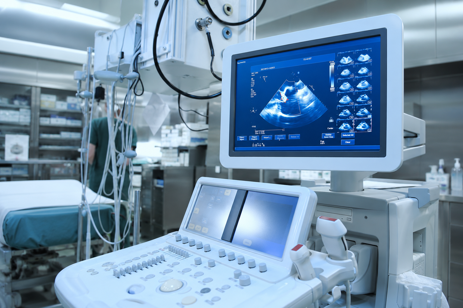
Ultrasound Scan in Thiruporur
Safe and Non-Invasive Imaging at AVR Scan Centre
An ultrasound scan is one of the most commonly performed diagnostic imaging examinations used to evaluate various organs, tissues, and structures within the body. Unlike CT scans and X-rays, ultrasound uses high-frequency sound waves to create real-time images and does not involve ionizing radiation.
Doctors may recommend an ultrasound scan for a wide range of clinical situations, including abdominal pain, pregnancy evaluation, pelvic conditions, thyroid disorders, kidney problems, gallbladder assessment, and vascular examinations.
Because ultrasound provides real-time imaging, it plays an important role in diagnosing, monitoring, and evaluating many medical conditions across different age groups.
At AVR Scan Centre, Thiruporur, ultrasound imaging services are available for patients seeking diagnostic evaluation based on their doctor's recommendation. Patients from Thiruporur, Kelambakkam, Padur, Navalur, Siruseri, Chengalpattu, and nearby OMR areas can conveniently access ultrasound services.
The specific type of ultrasound examination required depends on the symptoms, medical history, and clinical question that needs to be answered.
What Is an Ultrasound Scan?
An ultrasound scan, also known as sonography, is a diagnostic imaging examination that uses high-frequency sound waves to create images of structures inside the body.
Unlike X-rays and CT scans, ultrasound does not use radiation.
Instead, a small handheld device called a transducer sends sound waves into the body. These sound waves bounce off tissues and organs, and a computer converts the returning signals into real-time images.
Ultrasound imaging can be used to evaluate:
- Liver
- Gallbladder
- Kidneys
- Pancreas
- Spleen
- Thyroid gland
- Urinary bladder
- Female reproductive organs
- Pregnancy and fetal development
- Blood vessels
- Soft tissues
Because ultrasound provides live imaging, it is particularly useful for evaluating movement, blood flow, and organ function.
Ultrasound Scan in Thiruporur – When Might a Doctor Recommend One?
Doctors recommend ultrasound examinations for many different medical reasons.
The purpose of the scan depends on the area being examined and the symptoms being investigated.
Common reasons include:
- Abdominal pain
- Pelvic pain
- Pregnancy monitoring
- Kidney stone evaluation
- Thyroid swelling
- Breast lumps
- Gallbladder concerns
- Liver assessment
- Urinary symptoms
- Blood vessel evaluation
Ultrasound is often chosen because it is non-invasive, widely used, and capable of providing valuable diagnostic information without exposing patients to radiation.
What Types of Ultrasound Scans Are Available?
Ultrasound is not a single examination.
Different ultrasound studies are performed depending on the body part being evaluated.
Abdominal Ultrasound
Used to assess organs such as:
- Liver
- Gallbladder
- Pancreas
- Spleen
- Kidneys
Pelvic Ultrasound
Used to evaluate:
- Uterus
- Ovaries
- Urinary bladder
- Pelvic structures
Pregnancy Ultrasound
Used to monitor fetal growth and development during pregnancy.
Thyroid Ultrasound
Used to assess thyroid size, nodules, and other thyroid-related abnormalities.
Breast Ultrasound
May be used to evaluate breast lumps and selected breast-related concerns.
Doppler Ultrasound
Used to assess blood flow within arteries and veins.
Soft Tissue Ultrasound
Used for evaluating lumps, swelling, and soft tissue abnormalities.
How Does an Ultrasound Scan Work?
Ultrasound imaging uses high-frequency sound waves to create real-time images of organs and tissues inside the body. Unlike X-rays and CT scans, ultrasound does not use radiation.
During the examination, a trained healthcare professional places a handheld device called a transducer on the skin over the area being examined. A water-based gel is applied first to help the sound waves travel efficiently between the transducer and the body.
The transducer sends sound waves into the body and receives the echoes that bounce back from internal structures. A computer processes these echoes and converts them into live images displayed on a monitor.
Because ultrasound provides real-time visualization, doctors can assess:
What Doctors Can Assess with Ultrasound
- Organ structure
- Organ movement
- Blood flow
- Fluid collections
- Fetal development during pregnancy
- Soft tissue abnormalities
This ability to view structures dynamically makes ultrasound one of the most versatile diagnostic imaging tools available.
What Is a Pregnancy Ultrasound Scan?
Pregnancy ultrasound is one of the most commonly performed ultrasound examinations.
It is used to monitor fetal development and provide information about the progress of pregnancy.
Depending on the stage of pregnancy and the clinical requirement, ultrasound may help evaluate:
What a Pregnancy Ultrasound May Evaluate
- Fetal growth
- Fetal position
- Number of fetuses
- Placental location
- Amniotic fluid levels
- Fetal heartbeat
- General fetal development
Pregnancy scans are performed at different stages of pregnancy for different purposes. The timing and type of examination depend on the recommendations of the obstetrician or healthcare provider.
Ultrasound imaging plays an important role in prenatal care because it provides valuable information without exposing the mother or baby to ionizing radiation.
What Is the Difference Between 2D, 3D and 4D Ultrasound?
Many patients hear terms such as 2D, 3D, and 4D ultrasound and wonder what they mean.
All three use ultrasound technology, but they produce images differently.
2D Ultrasound
2D ultrasound creates standard flat images and remains the most commonly used ultrasound technique in medical practice.
It is widely used for:
- Pregnancy assessment
- Abdominal imaging
- Pelvic imaging
- Thyroid evaluation
- General diagnostic ultrasound
3D Ultrasound
3D ultrasound combines multiple image slices to create a three-dimensional representation of structures being examined.
It may provide enhanced visualization in selected clinical situations.
4D Ultrasound
4D ultrasound adds real-time movement to 3D imaging.
This allows moving three-dimensional images to be viewed during the examination.
The choice of examination depends on the clinical purpose and the recommendation of the healthcare professional.
What Is a Doppler Ultrasound?
A Doppler Ultrasound is a specialized ultrasound examination used to evaluate blood flow within arteries and veins.
Unlike routine ultrasound, which focuses primarily on anatomy and structure, Doppler imaging helps assess how blood moves through blood vessels.
Doctors may recommend Doppler studies when evaluating:
- Leg swelling
- Suspected vascular conditions
- Blood circulation concerns
- Varicose veins
- Arterial disorders
- Venous disorders
The examination provides valuable information regarding the direction and speed of blood flow.
Doppler studies are commonly used in vascular medicine, pregnancy care, and selected cardiovascular assessments.
What Can an Abdominal Ultrasound Evaluate?
An abdominal ultrasound is frequently recommended when doctors need to assess organs within the abdomen.
The examination may provide information about:
Liver
The liver can be evaluated for size, structure, and selected abnormalities.
Gallbladder
Ultrasound is commonly used to assess the gallbladder and investigate symptoms such as upper abdominal discomfort.
Kidneys
Kidney ultrasound may help evaluate kidney size, structure, and selected urinary tract concerns.
Pancreas
Parts of the pancreas may be visualized depending on patient anatomy and imaging conditions.
Spleen
The spleen can be assessed as part of a comprehensive abdominal examination.
Because abdominal symptoms can have many causes, ultrasound often serves as an important first-line imaging investigation.
What Is a Pelvic Ultrasound?
Pelvic ultrasound is used to evaluate structures located within the pelvic region.
For women, this examination may assess:
- Uterus
- Ovaries
- Endometrium
- Adnexal structures
- Pelvic organs
Doctors may recommend pelvic ultrasound when investigating:
- Pelvic pain
- Menstrual irregularities
- Fertility concerns
- Gynecological symptoms
- Follow-up of previously identified findings
Depending on the clinical situation, pelvic ultrasound may be performed using different approaches as determined by the healthcare provider.
What Is a Thyroid Ultrasound?
The thyroid gland is a small endocrine gland located in the front of the neck.
A thyroid ultrasound provides detailed images of the gland and surrounding structures.
Doctors may recommend thyroid ultrasound for:
- Thyroid swelling
- Neck lumps
- Thyroid nodules
- Thyroid enlargement
- Follow-up of known thyroid conditions
Ultrasound is particularly useful because it can provide detailed visualization of thyroid tissue without the use of radiation.
The examination is painless, non-invasive, and usually completed within a short period of time.
What Can a Kidney Ultrasound Show?
Kidney ultrasound is commonly performed when evaluating urinary or kidney-related symptoms.
The examination may help assess:
- Kidney size
- Kidney structure
- Urinary tract abnormalities
- Obstruction within the urinary system
- Selected kidney conditions
Doctors may recommend kidney ultrasound for patients experiencing:
- Flank pain
- Urinary symptoms
- Recurrent urinary infections
- Abnormal laboratory findings
- Kidney-related concerns
The scan provides important information that can assist healthcare professionals in evaluating kidney health.
What Can a Liver Ultrasound Show?
The liver performs many important functions within the body, including metabolism, detoxification, and nutrient processing.
Liver ultrasound may be recommended when doctors need to evaluate liver structure and appearance.
The examination may be used as part of the assessment for:
- Abnormal liver function tests
- Liver enlargement
- Fatty liver changes
- Abdominal symptoms
- Follow-up imaging
Ultrasound provides a non-invasive method of examining the liver and surrounding abdominal structures.
The findings are interpreted together with clinical information and laboratory investigations.
What Can a Gallbladder Ultrasound Evaluate?
Gallbladder ultrasound is one of the most frequently requested abdominal imaging examinations.
It is commonly recommended when patients experience symptoms such as:
- Upper abdominal discomfort
- Digestive symptoms
- Nausea after meals
- Suspected gallbladder disorders
Ultrasound can provide detailed images of the gallbladder and nearby structures.
Because ultrasound does not involve radiation and provides excellent visualization of the gallbladder, it is often the preferred initial imaging examination for gallbladder assessment.
How Should You Prepare for an Ultrasound Scan?
Preparation varies depending on the type of ultrasound examination being performed.
Different organs require different preparation methods to optimize image quality.
Abdominal Ultrasound
Some abdominal ultrasound examinations may require fasting before the scan.
Fasting can help improve visualization of certain abdominal organs.
Pelvic Ultrasound
Patients may be instructed to have a full bladder before the examination.
A full bladder can improve visualization of pelvic structures.
Pregnancy Ultrasound
Preparation requirements vary depending on the stage of pregnancy and the type of examination requested.
Thyroid Ultrasound
Generally, no special preparation is required.
Doppler Ultrasound
Preparation depends on the area being examined and the clinical indication.
Patients should follow the specific instructions provided when scheduling the appointment.
Is an Ultrasound Scan Safe?
Ultrasound is one of the most widely used imaging examinations in modern healthcare and has been used in clinical practice for several decades. Unlike imaging techniques that rely on ionizing radiation, ultrasound uses sound waves to create images of internal body structures.
Because it does not involve radiation exposure, ultrasound is commonly used across different age groups, including infants, children, adults, and pregnant women when clinically indicated.
Healthcare professionals may recommend ultrasound examinations for evaluating organs, blood vessels, soft tissues, and pregnancy-related conditions because of its safety profile and ability to provide real-time imaging.
Like any medical examination, ultrasound should be performed only when there is a valid clinical reason, and the type of scan should be determined by the referring healthcare provider.
Does an Ultrasound Scan Use Radiation?
One of the most common reasons doctors choose ultrasound imaging is that it does not use ionizing radiation.
Unlike:
- CT Scans
- X-Rays
- Fluoroscopy
- Mammography
ultrasound relies on high-frequency sound waves rather than radiation to produce images.
This makes ultrasound particularly useful for:
- Pregnancy monitoring
- Pediatric imaging
- Repeated follow-up examinations
- Soft tissue evaluation
- Abdominal imaging
- Pelvic imaging
Patients who are concerned about radiation exposure often ask whether ultrasound is a safer alternative. While the choice of imaging depends on the medical condition being investigated, ultrasound offers the advantage of radiation-free imaging when it is appropriate for the clinical question.


Is an Ultrasound Scan Painful?
Ultrasound examinations are generally painless and non-invasive.
During the scan, a transducer is moved over the skin surface after a small amount of gel has been applied. The gel helps improve contact between the transducer and the skin, allowing sound waves to travel effectively.
Most patients experience little more than mild pressure from the transducer.
Depending on the area being examined, the healthcare professional may need to apply slightly firmer pressure to obtain clear images. This can occasionally cause temporary discomfort, especially if the area being evaluated is already tender.
However, the examination itself is not typically painful and does not involve injections, surgical procedures, or tissue removal.
What Happens During an Ultrasound Scan?
Understanding the examination process can help patients feel more comfortable before their appointment.
The exact procedure depends on the type of ultrasound being performed, but the general process is similar for most examinations.
Registration and Preparation
Upon arrival, patients complete registration and any necessary preparation instructions are reviewed.
Positioning
The patient is positioned comfortably on the examination table.
The position varies depending on the body part being evaluated.
Application of Ultrasound Gel
A clear water-based gel is applied to the skin over the examination area.
The gel helps improve sound wave transmission and image quality.
Image Acquisition
The transducer is gently moved over the area being examined.
Real-time images appear on a monitor, allowing the healthcare professional to evaluate internal structures.
Completion of Examination
Once sufficient images have been obtained, the gel is removed and the examination is completed.
Most ultrasound scans are relatively quick, although examination time varies depending on the body part being assessed.
How Long Does an Ultrasound Scan Take?
The duration of an ultrasound examination depends on the complexity of the study and the area being evaluated.
Some examinations can be completed relatively quickly, while others require more detailed assessment.
Factors that influence scan duration include:
- Type of ultrasound
- Body part examined
- Clinical indication
- Patient anatomy
- Need for additional views
Most routine ultrasound examinations are completed within a reasonable appointment timeframe, although exact durations vary from patient to patient.
Patients should allow sufficient time for registration, preparation, image acquisition, and post-examination procedures.
What Are the Limitations of Ultrasound Imaging?
Although ultrasound is an extremely valuable diagnostic tool, every imaging examination has limitations.
Ultrasound provides excellent visualization of many organs and soft tissues, but there are situations where other imaging techniques may be more appropriate.
Some limitations include:
Image Quality Can Be Affected by Body Habitus
In certain patients, image quality may be reduced due to body composition and anatomical factors.
Gas Can Interfere with Imaging
Gas within the intestines can sometimes limit visualization of underlying structures.
Bone Blocks Ultrasound Waves
Because sound waves do not pass effectively through bone, ultrasound has limitations when evaluating structures located behind bony anatomy.
Not Every Condition Is Visible on Ultrasound
Certain conditions may require additional imaging such as CT, MRI, or other diagnostic examinations.
For this reason, doctors select imaging studies based on the specific clinical question being investigated.
What Happens After an Ultrasound Scan?
After the examination is completed, the images are reviewed and interpreted by the appropriate healthcare professional.
The findings are documented in a report that is provided to the referring doctor.
Your doctor evaluates the ultrasound findings together with:
- Symptoms
- Medical history
- Physical examination
- Laboratory investigations
- Previous imaging studies
The ultrasound report helps contribute to the overall diagnostic assessment.
Depending on the findings, the doctor may recommend:
- Observation
- Follow-up imaging
- Additional tests
- Specialist consultation
- Further treatment
Patients should discuss the results with their healthcare provider rather than attempting to interpret images independently.
Why Choose AVR Scan Centre for Ultrasound Scan in Thiruporur?
When searching for an Ultrasound Scan Centre in Thiruporur, patients often consider factors such as location, accessibility, availability of services, and comprehensive diagnostic support.
AVR Scan Centre provides ultrasound imaging services for patients requiring diagnostic evaluation based on their doctor's recommendation.
Convenient Location in Thiruporur
The centre is easily accessible for patients from:
- Thiruporur
- Kelambakkam
- Padur
- Navalur
- Siruseri
- Egattur
- Chengalpattu
- OMR Corridor
Wide Range of Ultrasound Examinations
Ultrasound services may be used for evaluating:
- Abdomen
- Pelvis
- Pregnancy
- Thyroid
- Kidneys
- Liver
- Gallbladder
- Soft tissues
- Blood vessels
Comprehensive Diagnostic Services
AVR Scan Centre also provides:
- MRI Scan 1.5T
- MultiSlice 3D CT Scan
- Omicron Covid CT Scan
- X-Ray
- 3D/4D Obstetrics
- Computerized ECG
- Echo Cardiogram
- SonoMammography
- Treadmill Test
- EEG
- PFT
- Lab Services
- Preventive Master Health Checkup
- Digital Mammogram
- OPG
Patient Guidance and Support
Patients can contact the centre before their appointment to confirm:
- Appointment availability
- Preparation instructions
- Referral requirements
- Examination details
What Should You Check Before Booking an Ultrasound Scan in Thiruporur?
Before scheduling an ultrasound examination, it is helpful to confirm a few important details.
Understand Why the Scan Has Been Recommended
Knowing the purpose of the examination can help you understand what information your doctor is seeking.
Ask About Preparation Requirements
Different ultrasound examinations require different preparation instructions.
Bring Previous Reports
If available, bring:
- Previous ultrasound reports
- CT reports
- MRI reports
- Blood test results
- Doctor referrals
Confirm Appointment Requirements
Contact the centre in advance to confirm appointment availability and preparation instructions.
