
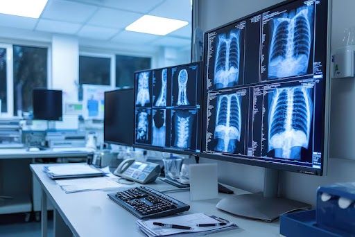
OPG X-Ray in Thiruporur
Panoramic Dental Imaging at AVR Scan Centre
If your dentist has recommended an OPG X-Ray in Thiruporur, you may want to understand what the examination shows, why it is recommended and how it differs from a regular dental X-ray.
An OPG (Orthopantomogram) is a panoramic dental X-ray that captures a broad image of the teeth, upper jaw, lower jaw and surrounding structures in a single scan. Unlike smaller dental X-rays that focus on a few teeth, an OPG provides a wider overview that can assist dentists in evaluating tooth position, wisdom teeth, jaw structures and treatment planning.
Dentists and oral healthcare professionals commonly use OPG imaging when assessing impacted teeth, orthodontic treatment, dental implants, jaw abnormalities and other dental concerns.
At AVR Scan Centre, Thiruporur, OPG services are available for patients requiring panoramic dental imaging and jaw assessment.
Patients from Thiruporur, Kelambakkam, Padur, Navalur, Siruseri, Chengalpattu and nearby OMR locations can conveniently access OPG imaging services.
What Is an OPG X-Ray?
An Orthopantomogram (OPG) is a two-dimensional panoramic dental X-ray that captures the entire mouth in a single image, including:
- Teeth
- Upper jaw (maxilla)
- Lower jaw (mandible)
- Jaw bones
- Wisdom teeth
- Temporomandibular joints (TMJ)
- Selected surrounding structures
Unlike intraoral dental X-rays where the detector is placed inside the mouth, an OPG uses a rotating imaging system that moves around the patient's head to create a panoramic image.
The examination provides a broad overview of dental and jaw anatomy and is often used as an initial imaging assessment in dentistry.
OPG X-Ray in Thiruporur – Where Can You Get an OPG Scan?
If you are searching for an OPG X-Ray in Thiruporur, AVR Scan Centre provides panoramic dental imaging services for patients requiring dental and jaw evaluation.
The centre serves patients from:
- Thiruporur
- Kelambakkam
- Padur
- Navalur
- Siruseri
- Egattur
- Chengalpattu
- Nearby OMR locations
Before visiting, patients may wish to confirm:
- OPG availability
- Appointment requirements
- Reporting timelines
- Preparation instructions
- Whether additional dental imaging has been requested
Why Might a Dentist Recommend an OPG?
An OPG may be recommended when a broad view of the teeth and jaws is required for diagnosis or treatment planning.
Depending on the clinical situation, panoramic imaging may assist with:
- Wisdom teeth evaluation
- Impacted teeth assessment
- Orthodontic planning
- Braces assessment
- Dental implant planning
- Jaw bone evaluation
- TMJ assessment
- Dental development assessment
- Pre-extraction evaluation
- General dental screening
Because the examination covers a larger area than many traditional dental X-rays, it can provide valuable information for treatment planning.
What Can an OPG X-Ray Show?
An OPG provides a broad panoramic view of structures within the mouth and jaws.
Teeth
The examination can show the position and arrangement of teeth throughout the upper and lower jaws.
Wisdom Teeth
OPG imaging is commonly used to assess wisdom teeth, including teeth that are impacted or have not erupted normally.
Impacted Teeth
The scan can help identify teeth that remain trapped beneath the gums or jawbone.
Jaw Bone
Panoramic imaging can provide an overview of the jaw bones and certain jaw-related abnormalities.
Orthodontic Assessment
An OPG is frequently used as part of orthodontic treatment planning to evaluate tooth position and development.
What Conditions Can an OPG Help Evaluate?
Depending on the reason for the examination, an OPG may contribute to the evaluation of:
- Impacted teeth
- Wisdom tooth positioning
- Jaw cysts
- Certain jaw abnormalities
- Dental development concerns
- Tooth positioning issues
- Orthodontic treatment planning
- Dental implant assessment
- TMJ-related concerns
- Selected bone abnormalities
The significance of any finding depends on clinical evaluation and professional interpretation.
How Does an OPG X-Ray Work?
During an OPG examination, the X-ray tube and detector rotate around the patient's head while images are captured.
The rotating system creates a panoramic image showing the entire mouth and jaws in a single view. Unlike traditional intraoral dental X-rays, no detector is placed inside the mouth during image acquisition.
This imaging technique allows dentists and oral healthcare professionals to evaluate multiple structures simultaneously.
What Happens During an OPG X-Ray?
An OPG is a quick, non-invasive dental imaging examination that usually takes only a few minutes to complete.
1. Registration and Verification
Before the scan, the imaging team may review:
- Patient details
- Dentist referral (if available)
- Previous dental imaging
- Relevant dental history
2. Removal of Metal Objects
You may be asked to remove:
- Earrings
- Neck chains
- Spectacles
- Hair accessories containing metal
- Removable dental appliances if advised
Metal objects can sometimes affect image quality.
3. Positioning
The radiographer will position your head correctly within the OPG unit.
Proper positioning is important because even small movements can affect image clarity.
4. Bite Block Placement
You may be asked to gently bite on a positioning guide to help align the teeth and jaws correctly.
5. Image Acquisition
The imaging arm rotates around your head while the panoramic image is captured.
The scan itself is usually completed within seconds.
6. Completion
Once imaging is complete, you can return to your normal activities immediately.
No recovery period is required.
How Should You Prepare for an OPG X-Ray?
One advantage of an OPG is that it generally requires very little preparation.
Before your appointment:
Remove Metal Objects
Metal items around the face and neck should be removed before imaging.
Inform the Imaging Team About Pregnancy
Because the examination uses X-rays, patients who are pregnant or may be pregnant should inform the healthcare team before the scan.
Bring Previous Dental Images
Previous dental X-rays or reports may help dentists compare findings when necessary.
Follow Any Dentist Instructions
If your dentist has provided specific instructions regarding imaging, follow those recommendations before the appointment.
Does an OPG X-Ray Use Radiation?
Yes.
An OPG uses a small amount of ionizing radiation to create panoramic dental images.
Like other dental X-ray examinations, OPG imaging follows radiation safety principles designed to minimize unnecessary exposure while obtaining the information needed for dental assessment.
The actual radiation dose can vary depending on:
- Equipment
- Imaging technique
- Patient size
- Examination requirements
Healthcare professionals recommend OPG imaging only when clinically appropriate.
Is an OPG X-Ray Safe?
OPG is a commonly performed dental imaging examination and is generally considered safe when used appropriately.
The benefits of obtaining important diagnostic information are weighed against the small radiation exposure associated with the examination.
Modern imaging systems are designed to:
- Optimize image quality
- Minimize radiation exposure
- Support safe imaging practices
Patients should always inform the healthcare team if they:
Important Information to Share Before the Examination
- Are pregnant
- May be pregnant
- Have questions about radiation exposure
Is an OPG X-Ray Painful?
No.
An OPG examination is generally painless and non-invasive.
There are:
- No injections
- No needles
- No surgical procedures
Most patients simply stand or sit comfortably while the machine rotates around the head.
Some individuals may find it slightly uncomfortable to remain still or maintain the required bite position, but the scan itself does not cause pain.
How Long Does an OPG X-Ray Take?
An OPG examination is usually completed quickly.
The actual image acquisition often takes less than a minute, while the overall appointment may take slightly longer due to:
- Registration
- Positioning
- Image verification
- Report processing
Most patients can complete the examination and continue their normal activities immediately afterward.
OPG vs Normal Dental X-Ray – What Is the Difference?
Although both examinations use X-rays, they provide different types of information.
| OPG (Panoramic X-Ray) | Intraoral Dental X-Ray |
|---|---|
| Captures the entire mouth and jaws | Focuses on a small area |
| Panoramic image | Detailed localized image |
| Detector remains outside the mouth | Detector placed inside the mouth |
| Useful for overall assessment | Useful for detailed tooth evaluation |
| Commonly used for treatment planning | Commonly used for specific dental problems |
Neither examination is universally better.
The most appropriate imaging test depends on the dental concern being evaluated.
OPG vs Dental CBCT – What Is the Difference?
An OPG and a CBCT scan provide different levels of information.
| OPG | Dental CBCT |
|---|---|
| Two-dimensional image | Three-dimensional imaging |
| Broad panoramic overview | Detailed 3D visualization |
| Commonly used as an initial assessment | Used when additional detail is required |
| Faster examination | More complex examination |
| Lower radiation exposure than many CBCT protocols | Radiation dose varies depending on protocol |
A CBCT is not automatically required for every dental problem.
Dentists select the most appropriate examination based on the clinical question.
Can an OPG Detect Wisdom Tooth Problems?
Yes.
An OPG is commonly used to evaluate wisdom teeth and their relationship to surrounding structures.
The examination may help assess:
- Erupted wisdom teeth
- Partially erupted wisdom teeth
- Impacted wisdom teeth
- Tooth angulation
- Developmental status
This information can assist dentists and oral surgeons when planning treatment.
Can an OPG Be Used for Braces and Orthodontic Treatment?
Yes.
Orthodontists frequently use panoramic imaging during treatment planning.
An OPG may provide information about:
- Tooth position
- Tooth development
- Eruption patterns
- Jaw relationships
- Impacted teeth
The examination is often combined with other clinical assessments and imaging when planning orthodontic treatment.
Can an OPG Be Used Before Dental Implants?
An OPG may be used as part of the initial assessment before dental implant treatment.
The panoramic image can provide an overview of:
- Existing teeth
- Jaw structures
- Bone appearance
- Treatment areas
However, some implant procedures may require additional imaging such as CBCT when detailed three-dimensional evaluation is necessary.
What Are the Limitations of an OPG X-Ray?
Although OPG imaging provides a valuable overview of the teeth and jaws, it has limitations.
These may include:
- Two-dimensional imaging only
- Less detail than some intraoral dental X-rays
- Limited soft tissue information
- Possible image distortion
- Overlapping structures
- Reduced ability to evaluate very small abnormalities
Because of these limitations, dentists may occasionally recommend additional imaging examinations.
Can an OPG Detect Oral Cancer?
An OPG is not a standalone test for diagnosing oral cancer.
While the examination may show certain jaw-related abnormalities, diagnosing oral cancer requires clinical assessment and may involve additional investigations.
If a suspicious lesion is identified, healthcare professionals may recommend:
- Clinical examination
- Specialist evaluation
- Additional imaging
- Biopsy when appropriate
A definitive diagnosis should never be based solely on an OPG image.
What Happens After an OPG X-Ray?
Once the panoramic image has been captured, the examination is complete and you can usually return to your normal activities immediately.
The OPG image is reviewed as part of your overall dental assessment. Dentists and oral healthcare professionals evaluate the findings together with:
- Your dental symptoms
- Clinical examination
- Medical and dental history
- Previous dental imaging
- Treatment requirements
An OPG provides a broad overview of the teeth and jaws, but the image should always be interpreted within the clinical context.
Additional investigations may sometimes be recommended if more detailed information is required.
Why Choose AVR Scan Centre for an OPG X-Ray in Thiruporur?
When searching for an OPG X-Ray in Thiruporur, patients often look for a diagnostic centre that offers convenient access to dental imaging and supports dentists with high-quality panoramic radiography.
Convenient Location for Thiruporur and OMR Residents
AVR Scan Centre serves patients from:
- Thiruporur
- Kelambakkam
- Padur
- Navalur
- Siruseri
- Egattur
- Chengalpattu
- Nearby OMR locations
Panoramic Dental Imaging Services
OPG imaging may be requested for:
- Wisdom tooth evaluation
- Orthodontic assessment
- Dental implant planning
- Tooth position assessment
- Jaw evaluation
- Pre-treatment dental imaging
Access to Multiple Diagnostic Services
AVR Scan Centre also provides:
- MRI Scan 1.5T
- MultiSlice 3D Spiral CT Scan
- Omicron COVID CT Scan
- Ultrasound Scan
- X-Ray
- 3D/4D Obstetric Ultrasound
- Computerized ECG
- Echo Cardiogram
- Treadmill Test
- EEG
- Pulmonary Function Test (PFT)
- Lab Services
- Preventive Master Health Checkup
- Digital Mammogram
- SonoMammography
Patient Guidance Before Imaging
Patients can contact the centre before visiting to confirm:
- OPG availability
- Appointment requirements
- Preparation instructions
- Reporting timelines
- Additional imaging requirements
Why Choose AVR Scan Centre for an OPG X-Ray in Thiruporur?
When searching for an OPG X-Ray in Thiruporur, patients often look for a diagnostic centre that offers convenient access to dental imaging and supports dentists with high-quality panoramic radiography.
Convenient Location for Thiruporur and OMR Residents
AVR Scan Centre serves patients from:
- Thiruporur
- Kelambakkam
- Padur
- Navalur
- Siruseri
- Egattur
- Chengalpattu
- Nearby OMR locations
Panoramic Dental Imaging Services
OPG imaging may be requested for:
- Wisdom tooth evaluation
- Orthodontic assessment
- Dental implant planning
- Tooth position assessment
- Jaw evaluation
- Pre-treatment dental imaging
Access to Multiple Diagnostic Services
AVR Scan Centre also provides:
- MRI Scan 1.5T
- MultiSlice 3D Spiral CT Scan
- Omicron COVID CT Scan
- Ultrasound Scan
- X-Ray
- 3D/4D Obstetric Ultrasound
- Computerized ECG
- Echo Cardiogram
- Treadmill Test
- EEG
- Pulmonary Function Test (PFT)
- Lab Services
- Preventive Master Health Checkup
- Digital Mammogram
- SonoMammography
Patient Guidance Before Imaging
Patients can contact the centre before visiting to confirm:
- OPG availability
- Appointment requirements
- Preparation instructions
- Reporting timelines
- Additional imaging requirements
What Should You Check Before Booking an OPG X-Ray in Thiruporur?
Before scheduling your OPG examination, it is useful to understand why the scan has been recommended and whether any preparation is required.
You may wish to confirm:
- The purpose of the examination
- Appointment availability
- Whether previous dental images are required
- Report delivery timelines
- Whether additional imaging has been requested by your dentist
You should also inform the imaging team if:
- You are pregnant or may be pregnant
- You have undergone recent dental procedures
- You have previous dental imaging available for comparison
Providing complete information helps ensure the examination is performed appropriately.
