
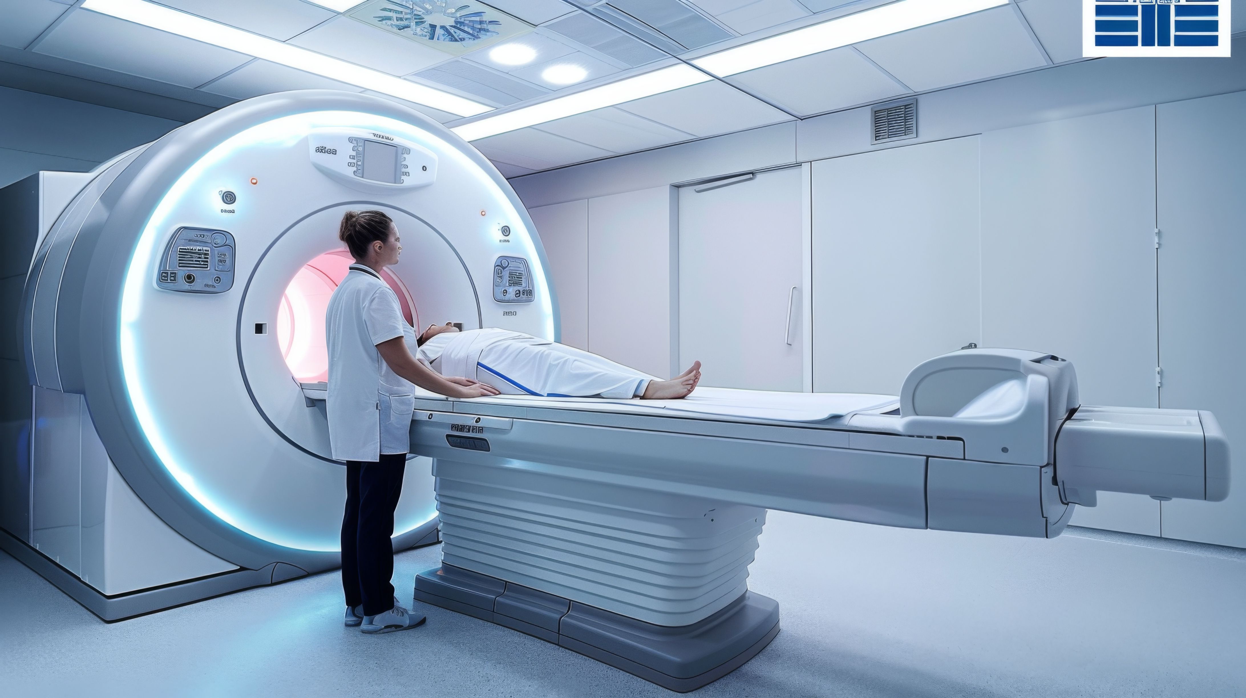
CT Scan in Thiruporur
CT Imaging at AVR Scan Centre
If your doctor has advised a CT Scan in Thiruporur, you may want to understand what the examination involves, why it has been recommended, and how it helps diagnose medical conditions.
A CT Scan (Computed Tomography Scan) is an advanced imaging examination that uses X-ray technology and computer processing to create detailed cross-sectional images of the body's internal structures. Modern Multi-Slice 3D CT scanners can produce highly detailed images of bones, blood vessels, organs, and tissues, helping doctors diagnose a wide range of medical conditions.
CT scans are widely used in emergency medicine, neurology, orthopedics, cardiology, oncology, pulmonology, and general medicine because they provide fast and detailed diagnostic information.
At AVR Scan Centre, Thiruporur, patients have access to Multi-Slice 3D CT Scan services for evaluating various medical conditions. Patients from Thiruporur, Kelambakkam, Padur, Navalur, Siruseri, Sholinganallur, Chengalpattu, and nearby OMR locations can conveniently access CT imaging services.
The specific CT examination required depends on your symptoms, clinical condition, and your doctor's recommendation.
What Is a CT Scan?
A CT Scan (Computed Tomography Scan) is an advanced diagnostic imaging examination that uses X-ray technology and computer processing to create detailed cross-sectional images of the body's internal structures. It provides doctors with clear and detailed views of bones, organs, blood vessels, and soft tissues, helping in the diagnosis and evaluation of various medical conditions.
Unlike a standard X-ray, which produces a single two-dimensional image, a CT scan captures multiple images from different angles. These images are processed and reconstructed into detailed slices, allowing doctors to examine internal structures more clearly and identify abnormalities with greater accuracy.
CT scans are commonly used to evaluate:
- Brain disorders
- Chest conditions
- Abdominal conditions
- Bone injuries
- Fractures
- Internal bleeding
- Blood vessel abnormalities
- Sinus disorders
- Spine conditions
Because CT imaging provides detailed visualization of both bones and internal organs, it has become one of the most commonly used diagnostic tools in modern medicine.
CT Scan in Thiruporur – Where Can You Get a CT Scan?
If you are searching for a CT Scan in Thiruporur, AVR Scan Centre provides Multi-Slice 3D CT imaging services for patients requiring advanced diagnostic evaluation.
CT scans may be recommended for a variety of medical conditions, ranging from injuries and trauma to neurological symptoms and internal organ evaluation.
Patients from the following areas frequently visit AVR Scan Centre:
- Thiruporur
- Kelambakkam
- Padur
- Navalur
- Siruseri
- Egattur
- Sholinganallur
- Chengalpattu
- OMR Region
Before your appointment, it is advisable to confirm CT scan availability, appointment requirements, and any preparation instructions associated with your examination.
AVR Scan Centre – Sholinganallur:
Address:
First Floor, No. 278/1 & 278/2, Commercial Building, Ramanyam Pushkar 2, Kalaignar Karunanidhi Salai, Sholinganallur – 600119.
MRI Service: 1.5T MRI Scan
Why Might a Doctor Recommend a CT Scan?
Doctors recommend CT scans when detailed imaging is required to investigate symptoms, confirm diagnoses, evaluate injuries, or assist with treatment planning.
Because CT scans provide rapid image acquisition and detailed anatomical information, they are commonly used in both routine and emergency medical situations.
A doctor may recommend a CT scan for:
- Persistent headaches
- Head injuries
- Stroke evaluation
- Sinus problems
- Chest pain
- Lung conditions
- Abdominal pain
- Kidney stones
- Bone fractures
- Sports injuries
- Internal injuries
- Tumor evaluation
- Blood vessel assessment
The reason for the examination depends on the patient's symptoms and the clinical question being investigated.
What Conditions Can a CT Scan Help Evaluate?
CT imaging is used to evaluate a wide variety of medical conditions affecting different parts of the body.
Neurological Conditions
CT Brain examinations may assist doctors in evaluating:
- Head injuries
- Stroke
- Brain bleeding
- Certain neurological abnormalities
Chest Conditions
CT Chest imaging may be used for:
- Lung abnormalities
- Chest infections
- Pulmonary conditions
- Evaluation of chest structures
Abdominal Conditions
CT Abdomen examinations may help evaluate:
- Kidney stones
- Abdominal pain
- Liver conditions
- Pancreatic disorders
- Intestinal abnormalities
Orthopedic Conditions
CT scans can provide detailed assessment of:
- Fractures
- Bone injuries
- Joint abnormalities
- Complex skeletal injuries
Vascular Conditions
Specialized CT studies may be used to evaluate blood vessels and circulatory structures when clinically indicated.
What Areas of the Body Can a CT Scan Examine?
CT scans can be performed on various parts of the body depending on the medical condition being investigated and the area that requires evaluation.
CT Brain
Provides detailed images of brain structures and may be used for neurological evaluation, head injuries, and other suspected brain conditions.
CT Sinus
Used to assess the sinus anatomy and evaluate sinus-related conditions and abnormalities.
CT Chest
Provides detailed images of the lungs and other structures within the chest, helping doctors evaluate various chest and pulmonary conditions.
CT Abdomen
Helps evaluate abdominal organs and internal structures, including the liver, kidneys, pancreas, and intestines.
CT Pelvis
Used to assess pelvic organs and surrounding anatomical structures when detailed imaging is required.
CT Spine
Provides detailed imaging of the spinal bones and related structures, helping evaluate fractures, injuries, and other spinal abnormalities.
CT Angiography
A specialized CT imaging examination used to evaluate blood vessels when recommended by a doctor based on the patient's symptoms and clinical condition.
What Is a Multi-Slice 3D CT Scan?
A Multi-Slice 3D CT Scan is an advanced form of CT imaging that captures multiple slices of data simultaneously.
This technology allows the scanner to acquire images rapidly while producing detailed cross-sectional and three-dimensional reconstructions of internal anatomy.
The benefits of Multi-Slice CT technology include:
- Faster image acquisition
- Detailed anatomical visualization
- Improved diagnostic information
- 3D image reconstruction capabilities
- Enhanced evaluation of bones, organs, and blood vessels
The exact imaging protocol depends on the body part being examined and the doctor's clinical requirements.
How Does a CT Scan Work?
CT imaging uses rotating X-ray equipment and advanced computer processing to create detailed images of structures inside the body.
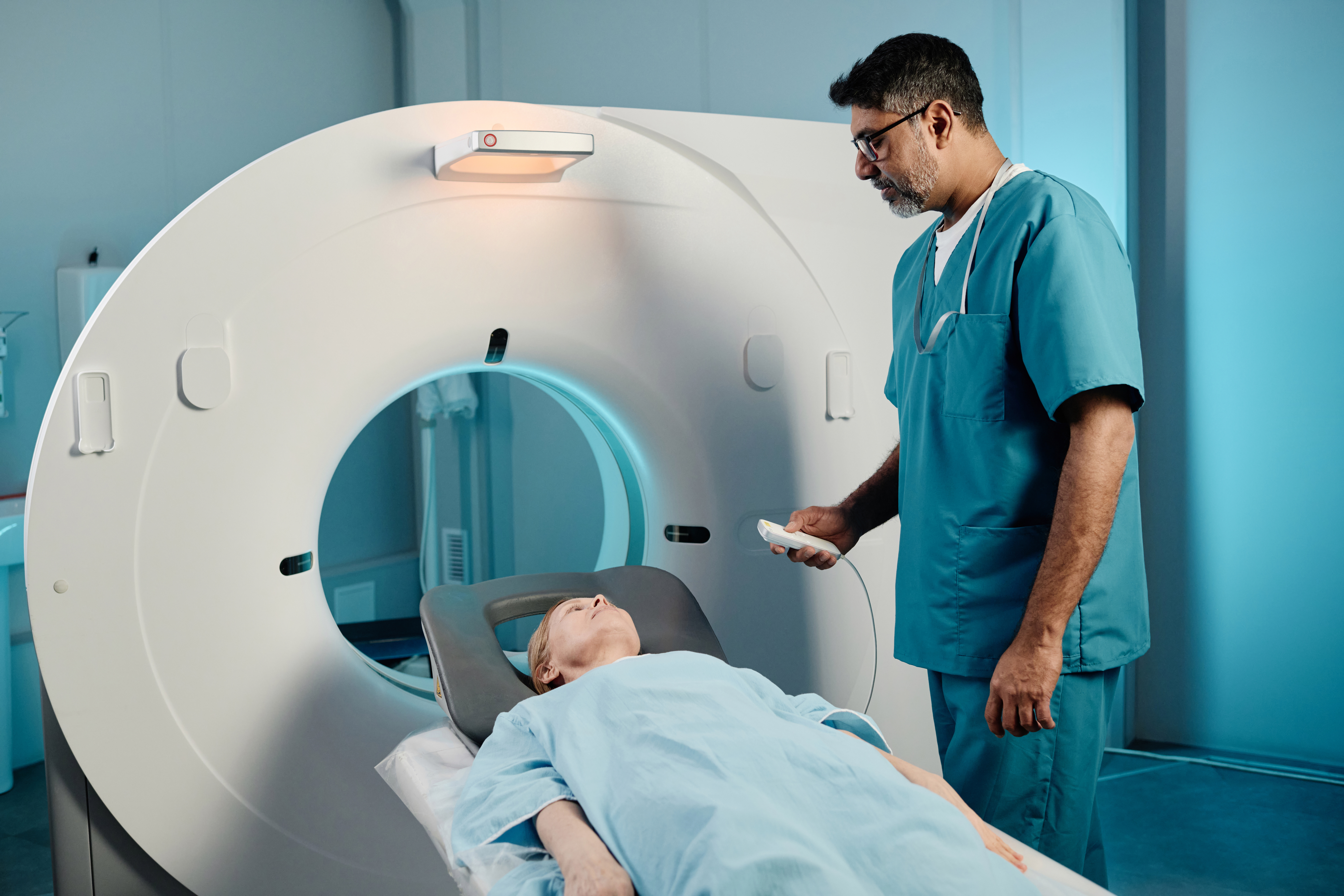
During the examination, the CT scanner rotates around the patient and captures multiple images from different angles. A computer processes these images and reconstructs them into detailed cross-sectional slices.
These slices can be viewed individually or combined to create three-dimensional representations of anatomical structures.
The resulting images help doctors evaluate bones, organs, tissues, blood vessels, and other internal structures and support the diagnosis of various medical conditions.
What Happens During a CT Scan?
Many patients are unfamiliar with CT imaging and may wonder what to expect during the examination.
Before the scan begins, the technologist will explain the procedure and provide any necessary instructions. Depending on the examination being performed, contrast material may or may not be required.
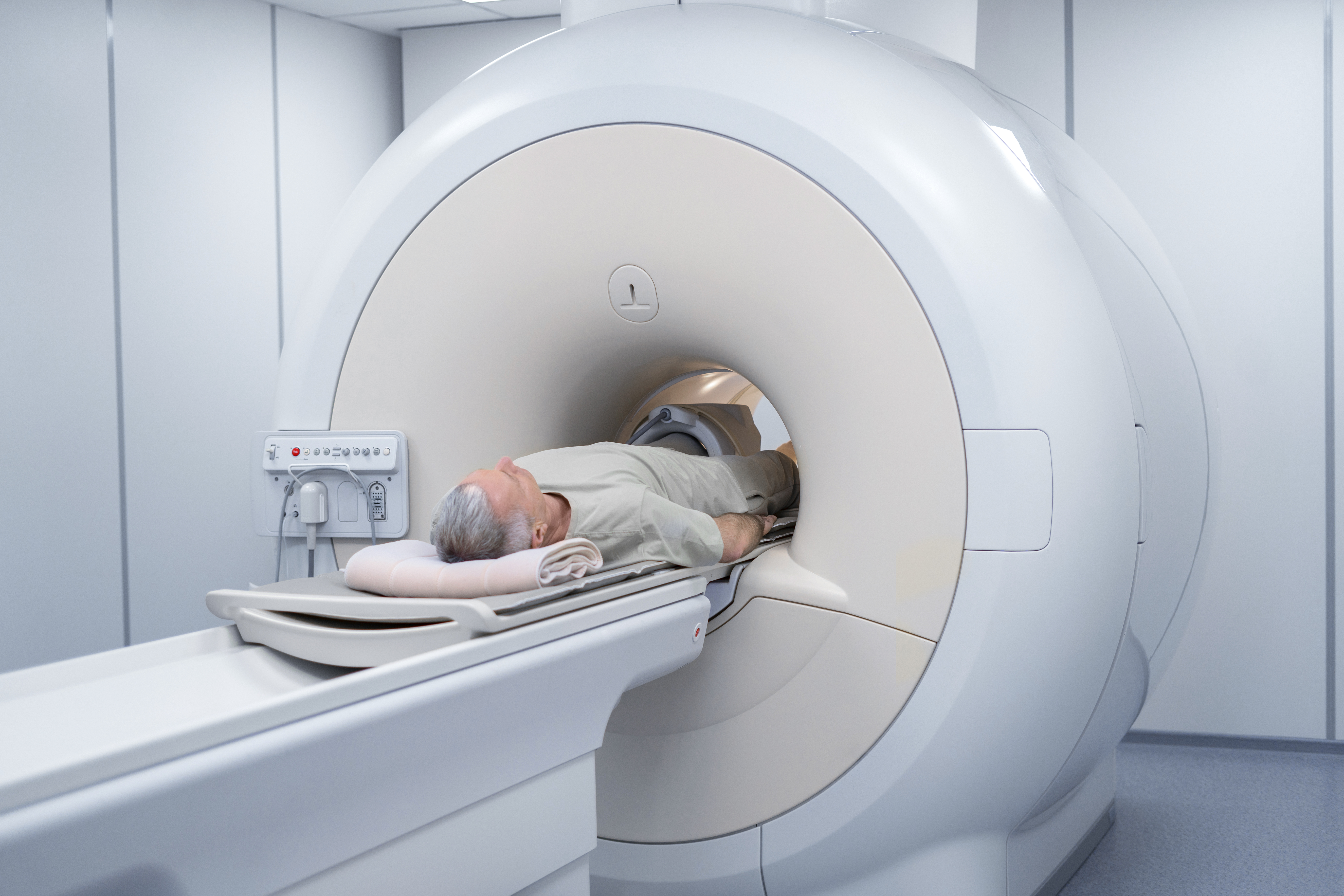
You will be positioned comfortably on the CT scanning table, which moves through the scanner opening during image acquisition.
The scanner captures images rapidly, and it is important to remain still to ensure image clarity.
Most CT examinations are completed within a relatively short period, although the total appointment time may vary depending on the type of study being performed.
The procedure is generally painless and non-invasive.
How Should You Prepare for a CT Scan?
Preparation depends on the type of CT examination being performed.
For many CT studies, little or no preparation may be required. However, some examinations involving contrast material may require specific instructions.
Patients should inform the imaging team about:
- Pregnancy
- Allergies
- Kidney disease
- Previous reactions to contrast material
- Medical conditions
- Current medications
Bringing previous imaging reports and referral documents may also assist in clinical evaluation.
Following any preparation instructions provided before your appointment can help ensure a smooth examination process.
Does a CT Scan Use Radiation?
A CT scan uses X-ray technology to create detailed cross-sectional images of the body. Unlike MRI, which uses magnetic fields and radiofrequency waves, CT imaging involves ionizing radiation.
Modern Multi-Slice CT scanners are designed to obtain high-quality diagnostic images while using optimized imaging protocols appropriate for the examination being performed. The amount of radiation exposure varies depending on the type of CT scan, the body area being examined, and the clinical requirements.
Doctors recommend CT examinations only when the diagnostic benefits outweigh the potential risks. The information obtained from a CT scan can play a critical role in diagnosing medical conditions, guiding treatment decisions, and monitoring patient health.
If you have concerns about radiation exposure, discuss them with your doctor or the radiology team before your examination.
Is a CT Scan Safe?
CT scans are commonly used in modern healthcare and are generally considered safe when performed for appropriate medical reasons.
Before the examination, the imaging team may review certain health factors that could affect the procedure, especially when contrast material is required.
Patients should inform the imaging team about:
- Pregnancy or possible pregnancy
- Kidney disease
- Previous reactions to contrast material
- Diabetes
- Current medications
- Known allergies
For most patients, CT imaging is completed without complications. The examination itself is non-invasive, and the scanning process is generally quick and well tolerated.
The decision to perform a CT scan should always be based on a doctor's assessment of the patient's symptoms and clinical needs.
Is a CT Scan Painful?
A CT scan is generally a painless and non-invasive examination.
During the examination, patients lie comfortably on the scanning table while images are acquired. The scanner does not touch the body, and the procedure does not involve needles unless intravenous contrast material is required for the specific examination.
Some patients may notice a warm sensation or metallic taste if contrast material is administered, but these sensations are usually temporary.
Most patients complete their CT examination comfortably and can return to normal activities shortly afterward unless instructed otherwise by their doctor.
Does Every CT Scan Require Contrast?
Not every CT examination requires contrast material.
Many CT scans can provide detailed diagnostic information without the use of contrast.
In certain cases, contrast-enhanced CT imaging may be recommended to provide clearer visualization of organs, blood vessels, tumors, inflammatory conditions, or specific anatomical structures.
Whether contrast material is required may depend on:
- The body part being examined
- The symptoms being investigated
- The medical condition being evaluated
- The specific diagnostic information required
The radiologist and referring physician will determine whether contrast-enhanced imaging is appropriate based on your clinical condition and the purpose of the examination.
CT Scan vs MRI Scan – What Is the Difference?
Although CT and MRI are both advanced diagnostic imaging examinations, they serve different clinical purposes.
CT scans use X-ray technology and are known for their speed and detailed visualization of bones, lungs, blood vessels, and various emergency conditions. MRI uses magnetic fields and radiofrequency waves and provides detailed images of soft tissues, including nerves, ligaments, spinal discs, muscles, and certain internal organs.
A CT scan is often chosen when rapid diagnosis is required, such as in trauma cases, suspected stroke, fractures, or internal injuries.
MRI is commonly selected when doctors require detailed evaluation of soft tissue structures and certain neurological conditions.
The most appropriate examination depends on your symptoms, medical history, and your doctor's clinical assessment.
Can a CT Scan Detect Fractures?
Yes. CT scans are highly effective for evaluating fractures and other skeletal injuries.
While standard X-rays are often used as the initial imaging examination for suspected fractures, a CT scan may be recommended when a more detailed assessment is required.
CT imaging can provide detailed visualization of:
- Complex fractures
- Facial bone injuries
- Pelvic fractures
- Spinal fractures
- Joint fractures
- Trauma-related bone injuries
The ability to produce cross-sectional and 3D images allows doctors to evaluate fracture patterns more accurately and plan treatment appropriately.
Can a CT Scan Detect Stroke?
CT Brain examinations play an important role in the early evaluation of suspected stroke.
In emergency situations, CT imaging is often one of the first investigations performed because it is fast, widely available, and can help identify certain types of brain abnormalities.
Doctors may use CT imaging to evaluate:
- Sudden weakness
- Speech difficulties
- Facial drooping
- Altered consciousness
- Suspected brain bleeding
- Acute neurological symptoms
The choice of imaging depends on the patient's condition and the clinical situation.
Can a CT Scan Detect Kidney Stones?
Yes. CT imaging is commonly used to evaluate suspected kidney stones and can provide detailed images of the urinary system.
Patients experiencing severe flank pain, blood in the urine, or symptoms suggestive of urinary tract obstruction may undergo CT imaging to help determine the presence, size, and location of kidney stones.
CT scans can also help doctors assess whether a stone is causing an obstruction or affecting surrounding structures.
Because of its detailed imaging capabilities, CT may be recommended when a thorough evaluation of suspected urinary tract stones is required.
Can a CT Scan Detect Lung Conditions?
CT Chest imaging provides detailed visualization of lung tissue and surrounding chest structures.
Doctors may recommend CT imaging when investigating:
- Persistent cough
- Lung infections
- Pulmonary abnormalities
- Chest symptoms
- Certain respiratory conditions
CT scans provide significantly more detail than a standard chest X-ray and can help evaluate conditions affecting the lungs, airways, and surrounding structures.
The interpretation of CT findings should always be performed in conjunction with the patient's symptoms and clinical examination.
What Are the Limitations of a CT Scan?
Although CT imaging is an important diagnostic tool, it may not be suitable for every medical situation.
CT scans may provide less soft tissue detail than MRI in certain circumstances. For example, MRI is often preferred when evaluating ligaments, spinal discs, nerves, and certain neurological conditions.
CT imaging also involves radiation exposure, which means examinations should be performed only when medically justified.
In some situations, additional imaging such as MRI, ultrasound, or laboratory investigations may be required to obtain a more complete understanding of a medical condition.
No single imaging test can answer every diagnostic question, which is why doctors select investigations based on individual patient needs.
What Happens After a CT Scan?
After your CT scan is completed, the images are processed and reviewed as part of the diagnostic evaluation. The examination itself is usually completed within a short period, allowing patients to return to their normal routine unless specific medical instructions have been provided.
The CT images are analyzed carefully and a report is prepared for the referring doctor. Your doctor will review the findings along with your symptoms, medical history, physical examination, and any previous investigations before discussing the results with you.
Depending on the reason for the scan, the findings may help:
- Confirm or rule out certain medical conditions
- Evaluate injuries or abnormalities
- Support treatment planning
- Monitor an existing condition
- Determine whether additional investigations are required
Patients should discuss the CT scan findings directly with their doctor, as imaging results must always be interpreted in the context of the patient's overall clinical condition.
Why Choose AVR Scan Centre for CT Scan in Thiruporur?
When looking for a CT Scan in Thiruporur, patients often consider factors such as diagnostic technology, accessibility, patient convenience, and the availability of comprehensive imaging services.
AVR Scan Centre provides Multi-Slice 3D CT Scan services designed to support accurate diagnostic evaluation for a wide range of medical conditions.
Advanced Multi-Slice CT Technology
The centre offers Multi-Slice 3D CT imaging capable of producing detailed cross-sectional and three-dimensional images that assist doctors in evaluating various conditions affecting the brain, chest, abdomen, bones, blood vessels, and other internal structures.
Convenient Location for Thiruporur and OMR Patients
AVR Scan Centre serves patients from:
- Thiruporur
- Kelambakkam
- Padur
- Navalur
- Siruseri
- Egattur
- Sholinganallur
- Chengalpattu
- OMR Corridor
The location provides convenient access for patients seeking advanced diagnostic imaging close to their community.
Guidance Throughout the Imaging Process
Patients often have questions regarding preparation, contrast administration, appointment requirements, and the examination procedure. The team provides information to help patients understand the process before their visit.
Comprehensive Diagnostic Services
In addition to CT imaging, AVR Scan Centre provides:
- MRI Scan 1.5T
- Ultrasound
- X-Ray
- 3D/4D Obstetrics
- Computerized ECG
- Echo Cardiogram
- SonoMammography
- Treadmill Test
- EEG
- PFT
- Lab Services
- Preventive Master Health Checkup
- Digital Mammogram
- OPG
This allows patients to access multiple diagnostic services through a single healthcare provider.
What Should You Check Before Booking a CT Scan in Thiruporur?
Before scheduling your CT examination, it is helpful to confirm a few important details to ensure a smooth and well-organized imaging experience.
Understand Why the CT Scan Has Been Recommended
Your doctor may recommend CT imaging to investigate symptoms, evaluate injuries, assess internal organs, or assist with treatment planning. Understanding the purpose of the examination can help you prepare appropriately.
Inform the Imaging Team About Medical Conditions
Patients should inform the imaging team if they have:
- Kidney disease
- Diabetes
- Previous reactions to contrast material
- Pregnancy or possible pregnancy
- Other ongoing medical conditions
Providing this information helps the imaging team determine whether any specific precautions or preparation may be required.
Bring Previous Reports
Previous imaging reports, including CT scans, MRI scans, X-rays, ultrasound reports, and relevant laboratory investigations, may provide useful information for comparison and clinical evaluation.
Confirm Preparation Requirements
Some CT examinations may require specific preparation, particularly when contrast-enhanced imaging is planned. Confirming the preparation instructions before your appointment can help avoid unnecessary delays.
Carry Your Referral Documents
Bringing your doctor's referral and relevant medical records can help the imaging team understand the clinical requirement and perform the appropriate CT examination.
Frequently Asked Questions About CT Scan in Thiruporur
A CT Scan (Computed Tomography Scan) is an advanced imaging examination that uses X-rays and computer technology to create detailed cross-sectional images of the body's internal structures.
AVR Scan Centre provides Multi-Slice 3D CT Scan services in Thiruporur for patients requiring diagnostic imaging of the brain, chest, abdomen, bones, blood vessels, and other parts of the body.
Yes. CT imaging uses X-ray technology, which involves ionizing radiation. The amount of exposure depends on the type of examination being performed.
CT scans are widely used in medical diagnosis and are considered safe when performed for appropriate clinical reasons under professional supervision.
Most CT examinations are completed within a short period. The exact duration depends on the type of scan and whether contrast material is required.
No. CT imaging is generally painless and non-invasive. Patients simply need to remain still while images are acquired.
Yes. CT imaging provides detailed evaluation of bones and is commonly used to assess complex fractures and skeletal injuries.
Yes. CT scans are frequently used to evaluate suspected kidney stones and determine their size and location.
CT scans use X-rays and are often preferred for trauma, fractures, and emergency imaging. MRI uses magnetic fields and is commonly used for soft tissue, nerve, ligament, and spinal evaluations.
No. Many CT examinations can be performed without contrast. Whether contrast is required depends on the medical condition being investigated and the doctor's recommendation.
Get Your CT Scan in Thiruporur at AVR Scan Centre
A CT scan provides detailed diagnostic information that helps doctors evaluate injuries, medical conditions, internal organs, bones, blood vessels, and many other structures throughout the body. Modern Multi-Slice 3D CT technology supports rapid and accurate imaging for a wide range of clinical applications.
At AVR Scan Centre, Thiruporur, patients can access Multi-Slice 3D CT Scan services supported by advanced imaging technology and patient-focused care.
Before visiting, it is advisable to confirm:
- Your required MRI examination
- Availability
- Appointment requirements
- Preparation instructions
- Any specific safety information you need to provide
AVR Scan Centre – Sholinganallur
First Floor, No. 278/1 & 278/2, Commercial Building, Ramanyam Pushkar 2, Kalaignar Karunanidhi Salai, Sholinganallur – 600119.
Contact AVR Scan Centre to enquire about the MRI examination recommended by your doctor and confirm appointment availability.
