

X-Ray in Thiruporur
Digital X-Ray Imaging at AVR Scan Centre
X-ray imaging remains one of the most commonly performed diagnostic examinations in healthcare. It provides doctors with a fast and effective way to evaluate bones, joints, the chest, and various parts of the body when symptoms require medical investigation.
Whether a patient is experiencing pain after an injury, persistent cough, joint discomfort, swelling, or other medical concerns, an X-ray may help doctors gather important information to support diagnosis and treatment planning.
Modern digital X-ray systems produce high-quality images that can be reviewed efficiently while helping healthcare professionals evaluate a wide range of conditions affecting the skeletal system and certain internal structures.
At AVR Scan Centre, Thiruporur, digital X-ray services are available for patients seeking diagnostic imaging based on their doctor's recommendation. Patients from Thiruporur, Kelambakkam, Padur, Navalur, Siruseri, Chengalpattu, and nearby OMR locations can conveniently access X-ray imaging services.
The type of X-ray required depends on the body part being examined and the clinical question that needs to be answered.
What Is an X-Ray?
An X-ray is a medical imaging examination that uses controlled amounts of ionizing radiation to create images of structures inside the body.
Different tissues absorb X-rays differently. Dense structures such as bones absorb more radiation and therefore appear differently on the image compared to soft tissues and air-filled spaces.
This difference allows doctors to visualize internal anatomy without surgery or invasive procedures.
X-ray imaging is commonly used to evaluate:
- Bones
- Joints
- Chest structures
- Lungs
- Spine
- Extremities
- Certain abdominal conditions
Because X-rays can be performed quickly and efficiently, they often serve as an important first-line diagnostic imaging examination.
X-Ray in Thiruporur – When Might a Doctor Recommend an X-Ray?
Doctors may recommend an X-ray for many different medical reasons.
The purpose of the examination depends on the symptoms, clinical findings, and area being investigated.
Some common reasons include:
- Suspected fractures
- Bone injuries
- Joint pain
- Persistent cough
- Chest symptoms
- Back pain
- Swelling
- Sports injuries
- Arthritis evaluation
- Follow-up of previous injuries
An X-ray often helps provide an initial assessment that guides further medical evaluation when necessary.
What Types of X-Rays Are Commonly Performed?
X-ray imaging can be used to examine many different parts of the body.
The type of examination depends on the clinical concern being investigated.
Chest X-Ray
One of the most frequently performed radiology examinations.
Used to evaluate structures such as:
- Lungs
- Heart silhouette
- Airways
- Chest cavity
Bone X-Ray
Used to evaluate:
- Fractures
- Bone alignment
- Bone abnormalities
- Injury-related changes
Joint X-Ray
Commonly performed for:
- Knee pain
- Shoulder pain
- Hip pain
- Ankle injuries
- Joint-related symptoms
Spine X-Ray
May be recommended for evaluating:
- Neck pain
- Back pain
- Spinal alignment
- Certain spinal abnormalities
Extremity X-Ray
Used for imaging:
- Hands
- Wrists
- Elbows
- Feet
- Ankles
- Legs
- Arms
Different examinations provide different types of information depending on the body part being assessed.
What Can a Chest X-Ray Show?
A Chest X-Ray is one of the most frequently requested imaging examinations.
Doctors may recommend a chest X-ray when evaluating respiratory symptoms or chest-related concerns.
The examination can help visualize:
Lungs
The lungs are assessed for abnormalities that may require further evaluation.
Airways
The visible airway structures can be evaluated as part of the examination.
Heart Size
The appearance of the heart silhouette may provide useful information.
Chest Structures
Various structures within the chest cavity can be visualized on X-ray imaging.
The interpretation of chest X-ray findings should always be performed by qualified healthcare professionals and considered alongside clinical information.
What Can a Bone X-Ray Evaluate?
Bones are among the structures most commonly examined using X-ray imaging.
A bone X-ray may help doctors evaluate:
- Fractures
- Bone alignment
- Joint relationships
- Bone healing
- Structural abnormalities
Because bone tissue appears clearly on X-ray images, radiography remains an important tool in orthopedic assessment and injury evaluation.
Patients experiencing trauma, falls, sports injuries, or unexplained bone pain may be referred for X-ray imaging depending on their symptoms and medical evaluation.
Can an X-Ray Detect Fractures?
One of the most common reasons for performing an X-ray is to evaluate suspected fractures.
When a bone breaks, changes in the bone structure may become visible on radiographic images.
Doctors often use X-rays to assess:
- Fracture location
- Bone alignment
- Degree of displacement
- Healing progress
However, it is important to understand that not every fracture is immediately visible on an initial X-ray.
In some situations, additional imaging studies or follow-up examinations may be recommended if symptoms persist despite normal initial findings.
How Does Digital X-Ray Technology Work?
Modern X-ray imaging has evolved significantly with the introduction of digital radiography.
Instead of using traditional film systems, digital X-ray technology captures images electronically.
During the examination, X-rays pass through the body and are detected by specialized digital detectors.
The resulting images are processed by a computer and displayed on a monitor for review.
Digital systems offer several advantages, including:
- Rapid image acquisition
- High-quality image visualization
- Efficient workflow
- Electronic image storage
- Easy image sharing when required
Digital radiography has become a widely used imaging method in modern diagnostic centres and hospitals.
Is a Digital X-Ray Different from a Traditional X-Ray?
The basic imaging principle remains the same.
Both traditional and digital X-ray systems use X-rays to create diagnostic images.
The primary difference lies in how the image is captured and processed.
Traditional systems relied on photographic film, whereas digital radiography uses electronic detectors and computer processing.
Digital imaging offers advantages such as:
- Faster image availability
- Improved image management
- Electronic storage
- Enhanced workflow efficiency
For patients, the examination experience is generally similar regardless of the image processing technology being used.
How Should You Prepare for an X-Ray?
In most cases, X-ray examinations require little or no special preparation. However, preparation may vary depending on the body part being examined and the reason for the investigation.
Before your appointment, it is helpful to follow any instructions provided by your doctor or imaging centre.
Patients may be asked to:
- Remove jewellery or metal objects
- Remove spectacles if they interfere with the imaging area
- Wear comfortable clothing
- Bring previous imaging reports if available
- Inform the healthcare team about pregnancy or possible pregnancy
Certain specialized X-ray examinations may require additional preparation, but routine diagnostic X-rays generally involve minimal preparation.
What Happens During an X-Ray Examination?
An X-ray examination is usually quick and straightforward.
When you arrive for your appointment, the imaging professional will explain the procedure and position you appropriately depending on the body part being examined.
For example:
Chest X-Ray
You may be asked to stand against the imaging detector while specific images are obtained.
Joint X-Ray
The affected joint will be positioned carefully to obtain images from different angles.
Spine X-Ray
Different positions may be required to visualize the spinal structures adequately.
Extremity X-Ray
The arm, hand, foot, ankle, or leg is positioned according to the examination requested.
During image acquisition, it is important to remain still because movement can reduce image clarity.
The imaging process itself usually takes only a few seconds, although the overall appointment may take longer due to positioning and preparation.
Does an X-Ray Use Radiation?
Yes. X-ray imaging uses controlled amounts of ionizing radiation to create diagnostic images.
The amount of radiation used depends on:
- Type of examination
- Body part being imaged
- Patient size
- Imaging technique
- Equipment used
Modern digital radiography systems are designed to obtain diagnostic images while using radiation doses that are appropriate for the clinical examination being performed.
Healthcare professionals follow established radiation safety principles to ensure that imaging is performed only when medically justified.
Is an X-Ray Safe?
X-ray examinations are widely used in modern healthcare and play an important role in diagnosing many medical conditions.
The decision to perform an X-ray is based on the potential diagnostic benefit compared with the small radiation exposure associated with the examination.
Medical imaging professionals follow radiation protection principles that include:
- Justification of the examination
- Appropriate image acquisition techniques
- Use of modern imaging technology
- Minimizing unnecessary exposure
Patients should inform the healthcare team if they are pregnant or think they may be pregnant before undergoing an X-ray examination.
When clinically appropriate, alternative imaging methods may sometimes be considered.
Is an X-Ray Painful?
No. A standard X-ray examination is generally painless.
The procedure does not involve injections, surgical instruments, or invasive techniques.
Some patients may experience temporary discomfort if an injured or painful body part needs to be positioned during the examination.
For example:
- Fractures
- Joint injuries
- Sports injuries
- Back pain
In these situations, the imaging professional works carefully to obtain the required images while minimizing discomfort whenever possible.
The X-ray itself is painless and usually completed quickly.
How Long Does an X-Ray Take?
Most routine X-ray examinations are completed within a relatively short period.
The exact duration depends on:
- Type of X-ray
- Number of images required
- Area being examined
- Patient positioning requirements
Simple examinations may take only a few minutes, while more complex studies requiring multiple views may take longer.
Patients should also allow time for:
- Registration
- Preparation
- Positioning
- Image review
The healthcare team will advise you if additional images are needed during the examination.
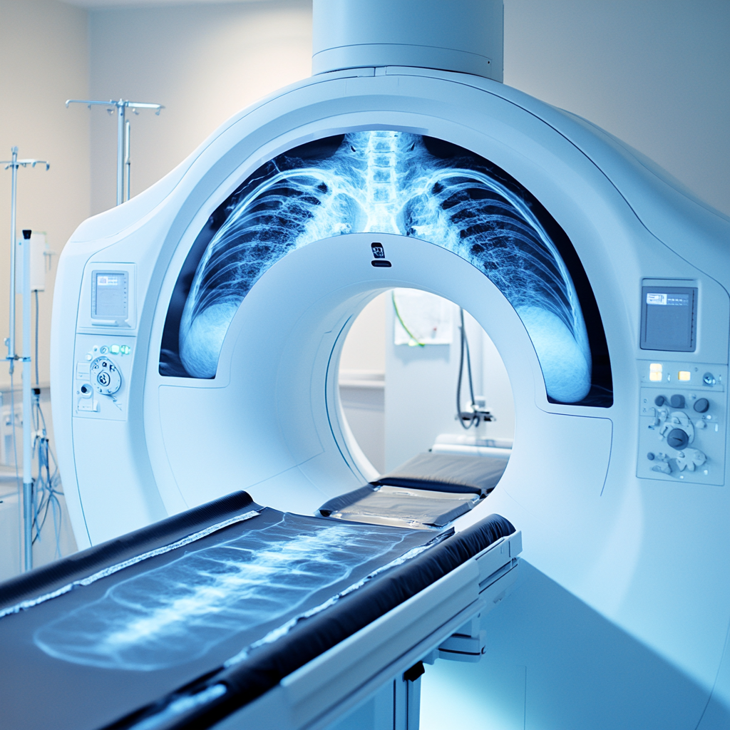

X-Ray vs CT Scan – What Is the Difference?
Although both X-ray and CT imaging use ionizing radiation, they provide different types of diagnostic information.
| X-Ray | CT Scan |
|---|---|
| Produces two-dimensional images | Produces detailed cross-sectional images |
| Often used as an initial imaging examination | Provides more detailed anatomical information |
| Commonly evaluates bones and chest structures | Can evaluate bones, organs, blood vessels and soft tissues |
| Faster and simpler examination | More complex imaging study |
| Often used for first-line assessment | Often used when additional detail is required |
Doctors select the most appropriate imaging examination based on the clinical situation and diagnostic requirements.
X-Ray vs MRI – What Is the Difference?
X-ray and MRI are fundamentally different imaging technologies.
X-Ray
Uses ionizing radiation to create images.
Commonly used for:
- Bones
- Joints
- Chest imaging
MRI
Uses magnetic fields and radiofrequency signals.
Commonly used for:
- Brain imaging
- Spine evaluation
- Soft tissues
- Ligaments
- Muscles
- Joints
MRI generally provides greater soft-tissue detail, while X-ray remains extremely valuable for evaluating skeletal structures and certain chest conditions.
X-Ray vs Ultrasound – What Is the Difference?
X-ray and ultrasound are used for different clinical purposes.
X-Ray
Provides images using ionizing radiation.
Often used for:
- Fractures
- Bone injuries
- Chest imaging
- Skeletal assessment
Ultrasound
Uses sound waves rather than radiation.
Commonly used for:
- Pregnancy imaging
- Abdomen evaluation
- Pelvic imaging
- Thyroid imaging
- Doppler studies
The most suitable examination depends on the symptoms and the information needed by the doctor.
Can an X-Ray Detect Arthritis?
X-rays are commonly used as part of the evaluation of joint-related symptoms.
The examination may help doctors assess:
- Joint alignment
- Joint space changes
- Bone abnormalities
- Degenerative changes
However, X-rays do not show every aspect of joint disease.
Depending on the clinical situation, additional imaging studies such as MRI or ultrasound may sometimes be recommended to provide further information.
The final diagnosis is based on clinical evaluation together with imaging findings.
Can an X-Ray Detect Lung Problems?
Chest X-rays are commonly used when doctors need to evaluate respiratory symptoms or chest-related concerns.
The examination may help visualize:
- Lungs
- Airways
- Chest cavity
- Heart silhouette
Doctors may recommend a chest X-ray when evaluating symptoms such as:
- Persistent cough
- Breathing difficulties
- Chest discomfort
- Respiratory infections
The X-ray findings must always be interpreted in conjunction with the patient's symptoms and medical history.
What Are the Limitations of X-Ray Imaging?
Although X-rays are an important diagnostic tool, they do have limitations.
Some conditions may not be fully evaluated using standard radiography alone.
Limited Soft Tissue Detail
Soft tissues are generally not visualized as clearly as they are with MRI or ultrasound.
Two-Dimensional Imaging
Standard X-rays provide two-dimensional images of three-dimensional structures.
Some Conditions May Require Additional Imaging
Doctors may recommend CT, MRI, ultrasound, or other examinations when more detailed evaluation is required.
Early Disease May Not Always Be Visible
Certain conditions may not produce obvious radiographic findings in their earliest stages.
For these reasons, imaging results are always interpreted together with clinical findings and other diagnostic information.
Can Pregnant Women Have an X-Ray?
Patients who are pregnant or may be pregnant should inform the healthcare team before any X-ray examination.
The healthcare provider will consider:
- Medical necessity
- Area being imaged
- Potential alternatives
- Clinical urgency
The decision regarding imaging during pregnancy should always be made by qualified healthcare professionals based on the individual clinical situation.
What Happens After an X-Ray?
Once the X-ray examination has been completed, the images are reviewed and processed for interpretation. The findings are then documented in a report that helps the referring doctor understand the condition being investigated.
The X-ray report is only one part of the diagnostic process. Your doctor will evaluate the findings together with:
- Your symptoms
- Medical history
- Physical examination
- Previous imaging studies
- Laboratory investigations if required
Depending on the clinical situation, the doctor may recommend:
- Observation and follow-up
- Medication or treatment
- Physiotherapy
- Orthopedic consultation
- Additional imaging such as CT, MRI, or Ultrasound
- Further diagnostic evaluation
Patients should always discuss the results with their healthcare provider to understand the significance of the findings and the next steps in management.
Why Choose AVR Scan Centre for X-Ray in Thiruporur?
When searching for an X-Ray Centre in Thiruporur, patients often look for accessibility, availability, quality imaging services, and a convenient diagnostic experience.
AVR Scan Centre provides digital X-ray imaging services for patients seeking diagnostic investigations recommended by their healthcare providers.
Convenient Location in Thiruporur
The centre is easily accessible for patients from:
- Thiruporur
- Kelambakkam
- Padur
- Navalur
- Siruseri
- Egattur
- Chengalpattu
- OMR Corridor
Digital X-Ray Imaging
Digital radiography helps produce high-quality images that support diagnostic evaluation across a wide range of medical conditions.
Multiple Diagnostic Services Under One Roof
AVR Scan Centre also provides:
- MRI Scan 1.5T
- MultiSlice 3D CT Scan
- Omicron Covid CT Scan
- Ultrasound
- 3D/4D Obstetrics
- Computerized ECG
- Echo Cardiogram
- SonoMammography
- Treadmill Test
- EEG
- PFT
- Lab Services
- Preventive Master Health Checkup
- Digital Mammogram
- OPG
Patient-Centered Diagnostic Support
Patients can contact the centre before their appointment to confirm:
- Appointment availability
- Preparation requirements
- Referral instructions
- Examination details
What Should You Check Before Booking an X-Ray in Thiruporur?
Before scheduling an X-ray examination, it is useful to confirm a few important details.
Understand the Purpose of the Examination
Knowing why your doctor has recommended an X-ray can help you understand the goal of the investigation and what information is being sought.
Bring Previous Reports
If available, bring:
- Previous X-rays
- CT reports
- MRI reports
- Ultrasound reports
- Medical records relevant to the condition
Previous studies can help healthcare professionals compare findings when necessary.
Inform the Team About Pregnancy
Women who are pregnant or may be pregnant should inform the imaging team before the examination.
Confirm Appointment Requirements
Some examinations may require appointments, while others may be available based on centre policies and schedules.
Contacting the centre in advance helps ensure a smooth experience.
Frequently Asked Questions About X-Ray in Thiruporur
An X-ray is a diagnostic imaging examination that uses controlled amounts of ionizing radiation to create images of structures inside the body.
Yes. X-ray imaging uses ionizing radiation to produce diagnostic images of bones, joints, chest structures, and other parts of the body.
No. X-ray examinations are generally painless. Some patients may experience mild discomfort if an injured area needs to be positioned during imaging.
Most routine X-ray examinations are completed within a short period, although the exact duration depends on the area being examined and the number of images required.
X-rays are commonly used to evaluate suspected fractures and other bone injuries. The findings are interpreted together with clinical information.
Chest X-rays may help doctors evaluate the lungs, airways, and chest structures when investigating respiratory symptoms.
An X-ray produces two-dimensional images, while a CT Scan creates detailed cross-sectional images that provide additional anatomical information.
X-rays are commonly used for bones and chest imaging, while MRI is often used for detailed evaluation of soft tissues, the brain, spine, and joints.
Most routine X-ray examinations require little or no preparation. Specific instructions depend on the type of examination being performed.
AVR Scan Centre provides digital X-ray imaging services for patients in Thiruporur and nearby areas along the OMR corridor.
Get Your X-Ray in Thiruporur at AVR Scan Centre
X-ray imaging continues to play an important role in modern healthcare by helping doctors evaluate a wide range of medical conditions affecting bones, joints, the chest, and other body structures.
Whether your doctor has recommended an X-ray for an injury, persistent symptoms, orthopedic assessment, respiratory concerns, or general diagnostic evaluation, understanding the purpose of the examination can help you prepare for your appointment.
AVR Scan Centre provides Digital X-Ray services in Thiruporur for patients seeking reliable diagnostic imaging and healthcare support.
Patients from Thiruporur, Kelambakkam, Padur, Navalur, Siruseri, Egattur, Chengalpattu, and surrounding OMR areas can conveniently access diagnostic imaging services at the centre.
Before your visit, contact the centre to confirm:
- Appointment availability
- Examination requirements
- Referral instructions
- Preparation guidelines
- Service availability
AVR Scan Centre – Thiruporur:
Address:
Ground Floor, No. 27, West Mada Street, Thiruporur, Tamil Nadu – 603110.
Contact AVR Scan Centre to enquire about the X-ray examination recommended by your doctor and confirm appointment availability.
