
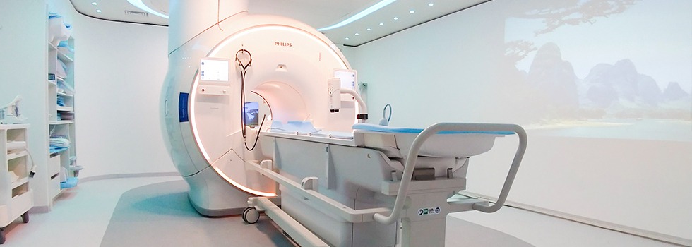
1.5T MRI at AVR Scan Centre
MRI Imaging at AVR Scan Centre
If your doctor has advised an MRI Scan in Thiruporur, understanding the purpose of the examination can help you feel more informed before your appointment. MRI, or Magnetic Resonance Imaging, is one of the most advanced diagnostic imaging techniques available today. It helps doctors visualize organs, tissues, nerves, joints, muscles, and other internal structures in remarkable detail without using ionizing radiation.
MRI plays an important role in modern healthcare because it provides detailed images that support diagnosis, treatment planning, and ongoing monitoring of many medical conditions. From evaluating headaches and neurological symptoms to assessing spinal disorders, sports injuries, joint pain, and soft tissue abnormalities, MRI has become a trusted diagnostic tool across multiple medical specialties.
At AVR Scan Centre, Thiruporur, patients have access to 1.5 Tesla MRI imaging services designed to assist doctors in obtaining high-quality diagnostic information. Patients from Thiruporur and nearby locations including Kelambakkam, Padur, Navalur, Siruseri, Egattur, Chengalpattu, and the OMR corridor can access MRI services conveniently.
The type of MRI examination recommended depends on your symptoms, clinical findings, and your doctor's assessment. Understanding how MRI works, why it may be recommended, and what to expect during the examination can help make the process more comfortable and stress-free.
What Is an MRI Scan?
MRI stands for Magnetic Resonance Imaging, a non-invasive diagnostic imaging technique used to create detailed images of structures inside the body. Unlike X-rays and CT scans, MRI does not use ionizing radiation. Instead, it uses powerful magnetic fields and radiofrequency waves to generate highly detailed images.
Doctors often recommend MRI when they need a closer look at soft tissues, muscles, nerves, ligaments, cartilage, internal organs, and other structures that may not be clearly using conventional imaging techniques.
MRI examinations are commonly used in:
- Neurology
- Orthopedics
- Spine Care
- Sports Medicine
- Gastroenterology
- Urology
- Gynecology
- Oncology
The images produced during an MRI examination help doctors understand the possible causes of symptoms and make informed decisions regarding treatment and further investigations.
MRI Scan in Thiruporur, Where Can You Get an MRI Scan?
If you are looking for an MRI Scan in Thiruporur, AVR Scan Centre provides MRI imaging services for patients seeking advanced diagnostic evaluation.
MRI scans are often performed when a doctor requires detailed information about the brain, spine, joints, nerves, muscles, or internal organs. Access to quality imaging services and proper patient guidance can help ensure a smooth diagnostic experience.
Patients from the following locations frequently visit AVR Scan Centre:
- Thiruporur
- Kelambakkam
- Padur
- Navalur
- Siruseri
- Egattur
- Sholinganallur
- Chengalpattu
- OMR Region
Before scheduling an MRI examination, it is advisable to confirm appointment requirements and any preparation instructions provided by the imaging centre or your doctor.
Why Might a Doctor Recommend an MRI Scan?
Doctors recommend MRI scans when detailed imaging is required to investigate symptoms, evaluate medical conditions, or support treatment planning.
Because MRI provides excellent soft tissue visualization, it is commonly used when symptoms involve the brain, spinal cord, nerves, muscles, joints, ligaments, tendons, and internal organs.
A doctor may recommend an MRI scan for:
- Persistent headaches
- Chronic neck pain
- Lower back pain
- Sciatica
- Joint pain
- Sports injuries
- Dizziness
- Seizures
- Numbness or tingling sensations
- Muscle weakness
- Nerve-related symptoms
- Suspected ligament injuries
- Evaluation of certain tumors or abnormalities
The specific reason for an MRI examination varies from patient to patient and should always be determined by a qualified healthcare professional.
What Conditions Can MRI Help Evaluate?
MRI is widely used because it provides detailed images of soft tissues and internal structures throughout the body.
Neurological Conditions
MRI may assist in evaluating:
- Stroke
- Brain tumors
- Multiple sclerosis
- Seizure disorders
- Chronic headaches
- Neurological abnormalities
Spine Disorders
MRI is commonly used to assess:
- Disc prolapse
- Herniated discs
- Sciatica
- Nerve compression
- Degenerative spinal changes
- Chronic neck pain
- Lower back pain
Sports and Orthopedic Injuries
MRI can help evaluate:
- Ligament tears
- Tendon injuries
- Meniscus tears
- Cartilage damage
- Muscle injuries
- Joint abnormalities
Abdominal and Pelvic Conditions
MRI may also be used in selected cases involving:
- Liver disorders
- Kidney abnormalities
- Pelvic conditions
- Prostate evaluation
- Reproductive system assessment
The interpretation of MRI findings should always be performed in conjunction with your symptoms, clinical examination, and medical history.
What Areas of the Body Can an MRI Scan Examine?
MRI can be performed on many different areas of the body depending on the medical condition being investigated.
MRI Brain
Used to evaluate neurological symptoms such as headaches, seizures, dizziness, stroke-related concerns, and other brain-related conditions.
MRI Spine
Provides detailed images of spinal discs, vertebrae, spinal cord, and nerve roots.
MRI Cervical Spine
Commonly recommended for neck pain, nerve compression, and cervical disc abnormalities.
MRI Lumbar Spine
Frequently used for lower back pain, disc prolapse, and sciatica.
MRI Knee
Helps evaluate ligaments, tendons, cartilage, menisci, and sports injuries.
MRI Shoulder
Useful for assessing rotator cuff injuries, tendon disorders, and shoulder instability.
MRI Pelvis
Provides detailed images of pelvic organs and surrounding structures.
MRI Abdomen
Used when detailed evaluation of abdominal organs is required.
What Is a 1.5 Tesla MRI Scanner?
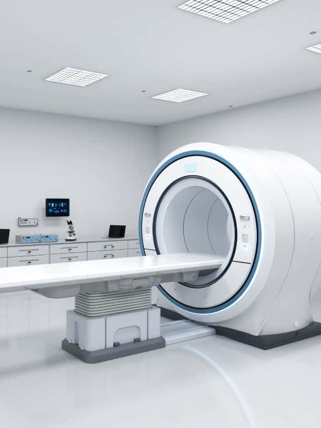
A 1.5 Tesla MRI scanner refers to the strength of the magnetic field used by the MRI system.
The 1.5T MRI platform is widely used around the world because it provides excellent image quality for a broad range of clinical applications. It is suitable for neurological imaging, spine imaging, orthopaedic examinations, musculoskeletal assessments, and many other diagnostic studies.
For many medical conditions, a 1.5 Tesla MRI scanner provides the detailed imaging information required for diagnosis and treatment planning.
How Does an MRI Scan Work?
MRI works by using a strong magnetic field and radiofrequency energy to generate detailed images of structures inside the body.
Hydrogen atoms present naturally within body tissues respond to the magnetic field and radiofrequency pulses produced by the scanner. These signals are detected and processed into detailed cross-sectional images.
The resulting images allow doctors to evaluate organs, tissues, nerves, muscles, joints, and other structures with a high level of detail.
What Happens During an MRI Scan?
Many patients feel anxious before their first MRI examination simply because they are unfamiliar with the procedure. Understanding what happens during the scan can help you feel more confident and prepared.
Before the examination begins, you will be asked a series of safety questions regarding your medical history, previous surgeries, implants, pacemakers, or any metallic objects that may affect MRI safety. This screening process is an important step in ensuring the examination can be performed safely.
You may be asked to remove items such as jewellery, watches, spectacles, hearing aids, mobile phones, belts, and other metallic accessories before entering the MRI room. Since MRI uses a powerful magnetic field, metal objects can interfere with both image quality and safety.
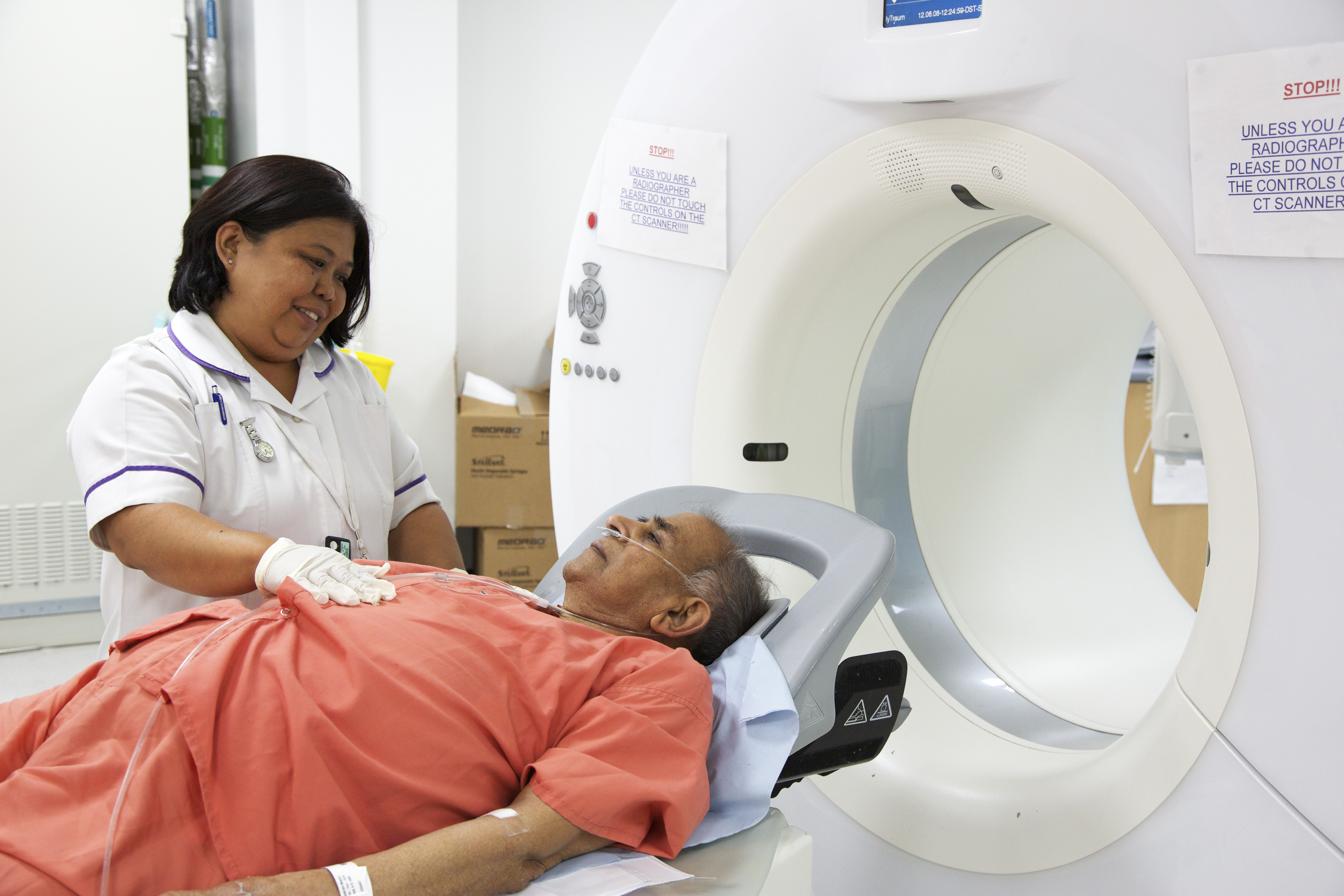
Once preparation is complete, you will be positioned comfortably on the MRI table. Depending on the area being examined, the technologist may use special coils that help capture detailed images. The table then moves into the MRI scanner, where images are acquired.
During the scan, you will hear repetitive tapping, knocking, or humming sounds. These sounds are a normal part of the MRI imaging process. Remaining as still as possible throughout the examination is important because movement can affect image clarity and may require images to be repeated.
The duration of the examination depends on the type of MRI being performed. Most MRI scans take between 20 and 60 minutes. After the scan is completed, you can usually return to your normal daily activities unless specific instructions have been provided by your doctor.
How Should You Prepare for an MRI Scan?
Most MRI examinations require minimal preparation, but following the instructions provided before your appointment can help ensure the scan proceeds smoothly.
The first and most important step is informing the MRI team about any medical devices, implants, or previous surgeries. Certain implants may require additional safety evaluation before the examination.
Patients should also inform the imaging team if they are pregnant, may be pregnant, or have a history of kidney problems, especially if a contrast-enhanced MRI has been requested.
Wearing comfortable clothing is generally recommended. Depending on the examination, you may be asked to change into a gown and remove all metallic accessories before entering the MRI suite.
Bringing previous imaging studies, medical records, and referral documents can also be helpful. Earlier MRI scans, CT scans, X-rays, or ultrasound reports may provide valuable information for comparison and assist the referring doctor in understanding changes over time.
Some MRI examinations involving contrast material may require additional preparation. If specific instructions apply to your examination, the imaging team will provide guidance before your appointment.
Does an MRI Scan Use Radiation?
One of the most common questions patients ask is whether MRI uses radiation.
The answer is no. MRI does not use ionizing radiation. Unlike conventional X-rays and CT scans, MRI relies on magnetic fields and radiofrequency waves to generate images. This allows doctors to obtain highly detailed images of internal structures without exposing patients to radiation during the examination.
Because of this characteristic, MRI has become an important imaging tool for evaluating a wide range of neurological, orthopedic, spinal, musculoskeletal, and internal medical conditions.
Although MRI does not use radiation, it should still only be performed when medically appropriate and recommended by a qualified healthcare professional.
Is an MRI Scan Safe?
MRI is considered a safe imaging examination for most patients when appropriate safety screening procedures are followed.
The examination does not involve radiation exposure and is routinely used around the world for diagnosing and monitoring a wide variety of medical conditions.
However, because MRI uses a powerful magnetic field, certain implanted devices and metallic objects require careful evaluation before scanning. Patients should always inform the MRI team if they have:
- Pacemakers
- Cochlear implants
- Surgical clips
- Artificial joints
- Neurostimulators
- Implanted pumps
- Metal fragments from previous injuries
- Any other implanted medical devices
The MRI team performs detailed safety screening before every examination to identify factors that may affect patient safety or image quality.
Patients who have questions about MRI safety should discuss their concerns with the healthcare team before the examination.
Is an MRI Scan Painful?
MRI is a non-invasive imaging procedure and is generally not painful.
The examination itself does not involve surgery or invasive treatment. Most patients experience little more than the need to remain still while images are acquired.
Some individuals with chronic back pain, neck pain, joint pain, or mobility issues may experience mild discomfort from lying in one position during the examination. If discomfort occurs, informing the MRI technologist can help ensure appropriate support and positioning.
The scanner may produce loud noises while images are being captured, but these sounds are completely normal. Hearing protection may be provided depending on the examination being performed.
For most patients, MRI is well tolerated and completed without significant discomfort.
Does Every MRI Scan Require Contrast?
Not every MRI examination requires contrast material.
In many situations, MRI provides sufficient diagnostic information without the use of contrast. However, certain medical conditions may require additional image enhancement to help doctors evaluate tissues, blood vessels, inflammation, or other abnormalities more clearly.
When contrast-enhanced imaging is considered beneficial, your doctor may recommend an MRI with contrast. The decision is based on the specific condition being investigated and the information required from the examination.
Before contrast administration, the healthcare team may review your medical history and assess factors that could influence the suitability of contrast use.
Whether contrast is required should always be determined by the referring doctor and radiology team based on the clinical indication.
MRI Scan vs CT Scan – What Is the Difference?
MRI and CT scans are both valuable diagnostic imaging tools, but they work in different ways and are used for different clinical purposes.
MRI uses magnetic fields and radiofrequency waves to generate images, whereas CT scans use X-ray technology to create cross-sectional images of the body.
MRI is particularly effective for evaluating soft tissues such as nerves, muscles, ligaments, tendons, cartilage, and the brain. CT scans are often preferred when rapid imaging is required or when detailed evaluation of bones and certain emergency conditions is needed.
| MRI Scan | CT Scan |
|---|---|
| Uses magnetic fields | Uses X-rays |
| No radiation exposure | Uses ionizing radiation |
| Excellent soft tissue detail | Excellent bone detail |
| Commonly used for brain, spine, nerves and joints | Commonly used for trauma and emergency imaging |
| Longer examination time | Faster examination time |
Neither examination is universally better than the other. The most appropriate imaging test depends on your symptoms, medical history, and the clinical question being investigated.
For many neurological, spinal, orthopedic, and soft tissue conditions, MRI is often chosen because of its ability to provide highly detailed images of internal structures.
Can MRI Detect a Slipped Disc?
A slipped disc, also known as a herniated or prolapsed disc, is one of the most common reasons doctors recommend MRI spine examinations.
MRI provides detailed visualization of spinal discs, vertebrae, nerve roots, and surrounding tissues. This allows doctors to determine whether a disc is bulging, herniated, or placing pressure on nearby nerves.
Patients experiencing persistent back pain, neck pain, leg pain, numbness, tingling sensations, or sciatica are often referred for MRI evaluation when disc-related problems are suspected.
Because MRI provides detailed images of both the spinal discs and surrounding nerve structures, it plays an important role in the assessment of many spinal conditions.
Can MRI Detect Nerve Compression?
Yes, MRI is one of the most commonly used imaging examinations for evaluating nerve compression and related spinal conditions.
Nerves can become compressed due to conditions such as disc prolapse, spinal canal narrowing, degenerative spine changes, or other structural abnormalities. When a nerve is compressed, symptoms may include pain, numbness, tingling sensations, muscle weakness, or discomfort that travels into the arms or legs.
MRI provides detailed images of the spinal cord, nerve roots, discs, and surrounding tissues, allowing doctors to assess whether a nerve is being affected and identify the possible cause of the symptoms.
For patients experiencing persistent nerve-related symptoms, MRI often provides valuable diagnostic information that helps guide further treatment decisions.
Can MRI Detect Ligament Injuries?
MRI is widely recognized as one of the most effective imaging techniques for evaluating ligament injuries, tendon injuries, cartilage damage, and other soft tissue abnormalities.
Unlike conventional X-rays, which primarily show bones, MRI provides detailed visualization of soft tissues throughout the body. This makes MRI particularly valuable when assessing sports injuries and joint-related conditions.
Doctors may recommend MRI to evaluate:
- ACL injuries
- PCL injuries
- MCL injuries
- Meniscus tears
- Rotator cuff injuries
- Tendon abnormalities
- Cartilage damage
- Muscle injuries
- Sports-related trauma
MRI helps healthcare professionals understand the extent of soft tissue injuries and supports treatment planning based on the findings.
Can MRI Detect Brain Disorders?
MRI plays an important role in the evaluation of many neurological conditions affecting the brain.
Because MRI provides highly detailed images of brain tissue, it is frequently used when patients experience symptoms such as persistent headaches, dizziness, seizures, balance problems, weakness, or other neurological concerns.
MRI Brain examinations may assist doctors in evaluating:
- Stroke
- Brain tumors
- Multiple sclerosis
- Certain infections
- Inflammatory conditions
- Structural abnormalities
- Neurological disorders
The information obtained from MRI imaging is combined with clinical findings, medical history, and other investigations to help doctors determine the most appropriate diagnosis and treatment plan.
What Are the Limitations of MRI?
Although MRI is an advanced diagnostic imaging tool, it is not suitable for every medical situation and has certain limitations.
One limitation is that MRI examinations typically take longer than CT scans. Remaining still during image acquisition is important because movement can affect image quality.
Certain implanted medical devices or metallic objects may also require additional safety assessment before MRI can be performed.
In some situations, doctors may recommend other imaging tests such as CT scans, ultrasound examinations, or X-rays if they are more appropriate for the condition being evaluated.
MRI also provides different information compared to other imaging modalities. Therefore, the choice of imaging depends on the specific clinical question and the information required by the treating doctor.
The most suitable imaging examination should always be determined by a healthcare professional based on individual patient needs.
What Happens After an MRI Scan?
After the MRI examination is completed, the images are reviewed as part of the diagnostic process.
The MRI images are carefully interpreted, and a report is prepared for the referring doctor. The doctor then reviews the findings alongside your symptoms, medical history, physical examination, and other investigations if necessary.
Depending on the purpose of the MRI examination, the results may help:
- Support diagnosis
- Monitor existing medical conditions
- Guide treatment planning
- Evaluate treatment response
- Determine whether additional investigations are needed
Patients are encouraged to discuss their MRI findings directly with their doctor, who can explain the significance of the results in the context of their overall health.
Why Choose AVR Scan Centre for MRI Scan in Thiruporur?
When searching for an MRI Scan in Thiruporur, patients often look for a diagnostic centre that combines advanced imaging technology, accessibility, patient-focused care, and a wide range of diagnostic services.
AVR Scan Centre is committed to supporting patients and healthcare professionals with reliable diagnostic imaging services designed to assist clinical decision-making.
Advanced 1.5 Tesla MRI Technology
AVR Scan Centre offers 1.5 Tesla MRI imaging, providing detailed diagnostic images that assist doctors in evaluating neurological, spinal, orthopaedic, musculoskeletal, and internal medical conditions.
Convenient Location for Thiruporur and OMR Residents
Located in Thiruporur, the centre serves patients from nearby locations including:
- Kelambakkam
- Padur
- Navalur
- Siruseri
- Egattur
- Chengalpattu
- Sholinganallur
- OMR Corridor
This accessibility helps patients receive diagnostic imaging closer to home.
Patient-Focused Experience
Many patients feel anxious before undergoing an MRI examination. The team at AVR Scan Centre aims to provide clear guidance regarding preparation, safety procedures, and appointment requirements to help patients feel informed throughout the process.
Comprehensive Diagnostic Services Under One Roof
In addition to MRI services, AVR Scan Centre provides:
- MultiSlice 3D Spiral CT Scan
- Ultrasound
- X-Ray
- 3D/4D Obstetrics
- Computerized ECG
- Echo Cardiogram
- SonoMammography
- Treadmill Test
- EEG
- PFT
- Lab Services
- Preventive Master Health Checkup
- Digital Mammogram
- OPG
This comprehensive range of services supports patients who may require multiple diagnostic investigations as part of their healthcare journey.
What Should You Check Before Booking an MRI Scan in Thiruporur?
Booking an MRI examination is generally straightforward, but confirming a few important details beforehand can help ensure a smooth experience.
Understand Why the MRI Has Been Recommended
Your doctor recommends MRI based on your symptoms and medical condition. Understanding the purpose of the examination helps you prepare and know what information is being evaluated.
Inform the MRI Team About Implants
Always inform the imaging team if you have:
- Pacemakers
- Cochlear implants
- Artificial joints
- Surgical clips
- Implanted medical devices
- Metal fragments
This information is important for MRI safety screening.
Carry Previous Reports
Previous MRI scans, CT scans, X-rays, ultrasound reports, and medical records may assist your doctor when comparing findings over time.
Confirm Preparation Instructions
Although many MRI examinations require minimal preparation, some studies may involve specific instructions, particularly if contrast-enhanced imaging has been requested.
Bring Your Doctor's Referral
Carrying your referral documents and medical records helps ensure the correct examination is performed according to your doctor's recommendation.
Frequently Asked Questions About MRI Scan in Thiruporur
MRI (Magnetic Resonance Imaging) is an advanced diagnostic imaging examination that uses magnetic fields and radiofrequency waves to create detailed images of organs, tissues, nerves, muscles, joints, and other structures inside the body without using ionizing radiation.
AVR Scan Centre provides MRI imaging services in Thiruporur for patients who require detailed diagnostic evaluation of the brain, spine, joints, muscles, nerves, and internal organs.
No. MRI does not use ionizing radiation. Instead, it uses magnetic fields and radiofrequency energy to generate diagnostic images.
MRI is generally considered safe for most patients when appropriate safety screening procedures are followed. Patients with pacemakers, implants, or certain medical devices should inform the MRI team before the examination.
The duration of an MRI examination varies depending on the body part being scanned and the complexity of the study. Most MRI examinations typically take between 20 and 60 minutes.
No. MRI is a non-invasive examination and is generally painless. Most patients simply need to remain still while images are being acquired.
MRI is commonly used to evaluate conditions affecting the brain, spine, nerves, muscles, joints, ligaments, tendons, and internal organs. Your doctor will recommend an MRI when detailed imaging is needed for diagnosis or treatment planning.
No. Many MRI examinations can be performed without contrast material. Whether contrast is required depends on the condition being investigated and the information needed from the scan.
MRI uses magnetic fields and radiofrequency waves, while CT scans use X-rays. MRI is particularly useful for evaluating soft tissues such as the brain, spine, nerves, muscles, and joints, whereas CT scans are often preferred for certain emergency conditions and bone-related evaluations.
Before an MRI examination, inform the imaging team about any implants, pacemakers, surgeries, or medical devices. Remove metallic objects and follow any preparation instructions provided by your doctor or the imaging centre.
Get Your MRI Scan in Thiruporur at AVR Scan Centre
An MRI scan is one of the most advanced diagnostic imaging examinations available today, helping doctors evaluate a wide range of neurological, orthopaedic, spinal, muscular, and internal medical conditions. By providing highly detailed images of the body's internal structures, MRI supports accurate diagnosis, treatment planning, and ongoing medical care.
Whether your doctor has recommended an MRI Brain, MRI Spine, MRI Knee, MRI Shoulder, MRI Pelvis, MRI Abdomen, or another MRI examination, understanding the purpose of the scan and following the recommended preparation instructions can help ensure a smooth imaging experience.
At AVR Scan Centre, Thiruporur, we are committed to providing advanced 1.5 Tesla MRI imaging services for patients across Thiruporur and surrounding areas. Our goal is to support patients and healthcare professionals with accessible diagnostic imaging services in a comfortable and professional environment.
Before your appointment, it may be helpful to confirm:
- MRI scan availability
- Appointment requirements
- Preparation instructions
- Whether contrast material is required
- Previous imaging reports to bring
- Any additional instructions recommended by your doctor
AVR Scan Centre – Thiruporur
First Floor, No. 278/1 & 278/2, Commercial Building, Ramanyam Pushkar 2, Kalaignar Karunanidhi Salai, Sholinganallur – 600119.
If your doctor has advised an MRI examination, AVR Scan Centre is here to assist you with advanced diagnostic imaging services and appointment guidance.
Contact AVR Scan Centre to enquire about the MRI examination recommended by your doctor and confirm appointment availability.
