

SonoMammography in Thiruporur
Breast Ultrasound at AVR Scan Centre
If your doctor has recommended a SonoMammography in Thiruporur, you may want to understand what the examination is, why it is performed, what it can detect, whether it is painful, how to prepare and what happens during the procedure.
SonoMammography, commonly known as a Breast Ultrasound, is a non-invasive imaging examination that uses sound waves to create images of breast tissue. It is frequently used to evaluate breast symptoms, investigate abnormalities found during a physical examination or mammogram, and provide additional information about breast tissue.
Unlike mammography, which uses low-dose X-rays, SonoMammography uses ultrasound technology and does not expose patients to radiation.
At AVR Scan Centre, Thiruporur, SonoMammography services are available for patients requiring breast imaging and diagnostic evaluation as advised by their healthcare provider.
Patients from Thiruporur, Kelambakkam, Padur, Navalur, Siruseri, Chengalpattu and nearby OMR locations can conveniently access breast ultrasound services.
What Is SonoMammography?
SonoMammography is a breast imaging examination performed using ultrasound technology.
The test uses high-frequency sound waves to create detailed images of breast tissue. A handheld ultrasound transducer is placed on the skin over the breast area, and the reflected sound waves are converted into images displayed on a monitor.
Doctors may recommend SonoMammography when they need additional information about breast tissue that cannot be obtained through physical examination alone.
The examination may help assess:
- Breast lumps
- Breast pain
- Breast swelling
- Breast cysts
- Dense breast tissue
- Breast abnormalities detected during screening
- Changes in breast tissue
Because ultrasound provides real-time imaging, it is commonly used as part of breast health evaluation.
SonoMammography in Thiruporur – Where Can You Get a Breast Ultrasound?
If you are searching for SonoMammography in Thiruporur, AVR Scan Centre provides breast ultrasound imaging services for patients requiring diagnostic breast assessment.
The centre serves patients from:
- Thiruporur
- Kelambakkam
- Padur
- Navalur
- Siruseri
- Egattur
- Chengalpattu
- OMR Corridor
AVR Scan Centre – Thiruporur:
Ground Floor, No. 27, West Mada Street, Thiruporur, Tamil Nadu – 603110
Before visiting, patients may wish to confirm:
- Breast ultrasound availability
- Appointment requirements
- Doctor referral requirements
- Previous imaging reports to bring
- Additional breast investigations requested by the doctor
Why Might a Doctor Recommend SonoMammography?
Doctors recommend SonoMammography for various breast-related concerns and diagnostic evaluations.
The examination may be requested when evaluating:
- Breast lumps
- Breast pain
- Localized breast swelling
- Changes in breast tissue
- Nipple-related symptoms
- Abnormal mammogram findings
- Dense breast tissue
- Follow-up assessment of previously identified findings
The exact reason for the examination depends on the patient's symptoms, medical history and clinical findings.
What Can SonoMammography Show?
SonoMammography provides detailed images of breast tissue and may help healthcare professionals evaluate a variety of breast-related findings.
Breast Lumps
Ultrasound can help evaluate the characteristics of breast lumps detected during examination or screening.
Breast Cysts
The examination may help distinguish fluid-filled cysts from certain other types of breast findings.
Dense Breast Tissue
In some situations, breast ultrasound may provide additional information when breast tissue is dense.
Breast Tissue Changes
The examination may help evaluate certain changes within breast tissue.
Follow-Up Imaging
SonoMammography may be used as part of ongoing monitoring when follow-up imaging is recommended.
What Conditions Can SonoMammography Help Evaluate?
SonoMammography is used as part of the assessment of various breast-related symptoms and abnormalities.
Depending on the clinical situation, the examination may contribute to the evaluation of:
- Breast cysts
- Breast lumps
- Dense breast tissue
- Localized breast abnormalities
- Breast pain
- Inflammatory breast changes
- Certain benign breast conditions
- Follow-up of previously identified breast findings
The interpretation of breast ultrasound findings should always be performed alongside clinical examination and other relevant investigations.
How Does SonoMammography Work?
SonoMammography uses ultrasound technology to create images of breast tissue.
During the examination, a transducer emits sound waves that travel into the breast tissue.
When these sound waves encounter different tissue structures, they reflect back toward the transducer.
The ultrasound system processes these returning echoes and converts them into real-time images that can be viewed on a monitor.
Because ultrasound uses sound waves rather than radiation, it is considered a non-invasive imaging technique widely used in medical diagnostics.
What Happens During a SonoMammography Examination?
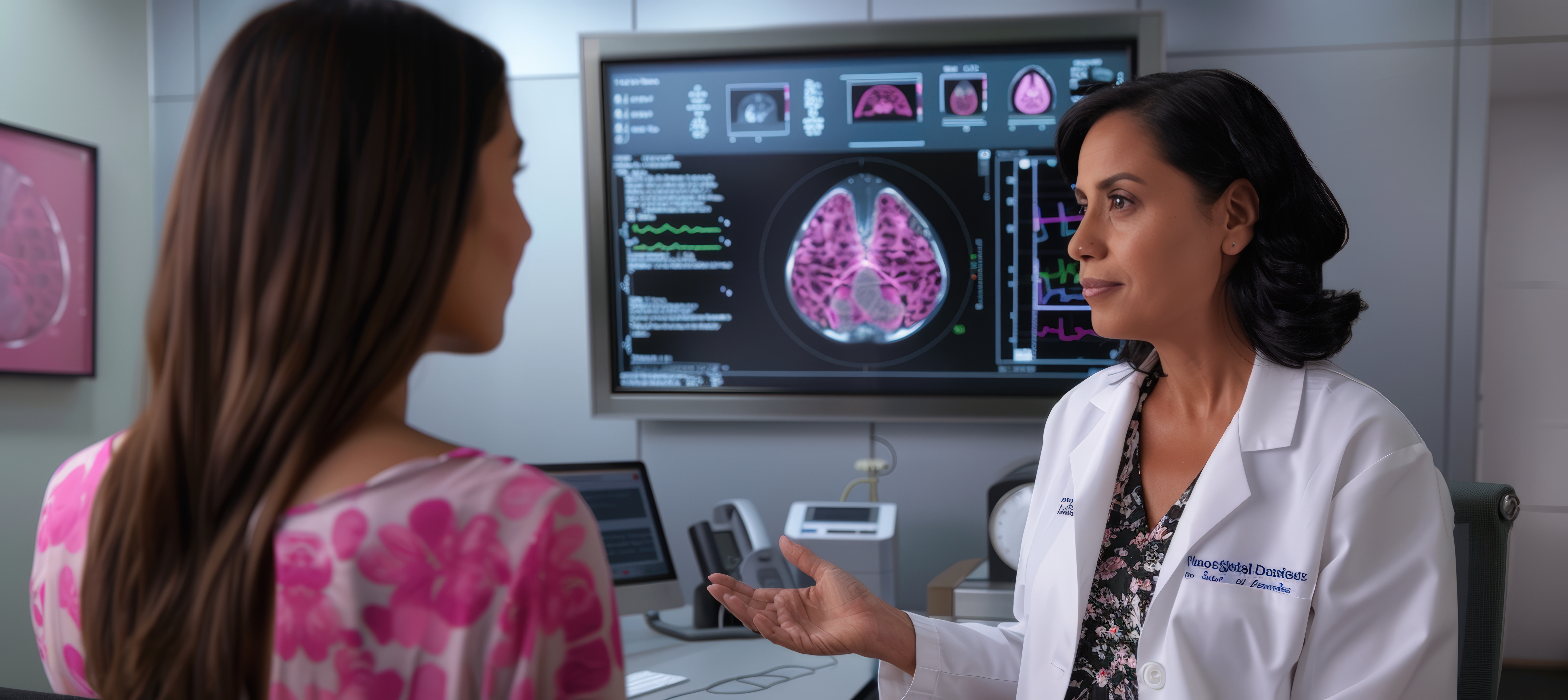
A SonoMammography examination is generally performed while the patient lies comfortably on an examination couch.
A clear water-based gel is applied to the skin of the breast area to improve the transmission of ultrasound waves.
The healthcare professional then gently moves the ultrasound transducer across the breast and surrounding areas to obtain images from different angles.
During the examination, additional images may be taken if specific areas require closer evaluation.
The procedure is generally well tolerated and does not require injections or recovery time.

How Should You Prepare for SonoMammography?
One of the advantages of SonoMammography is that it generally requires minimal preparation.
Most patients can continue their usual daily routine before the examination unless their doctor provides specific instructions.
Before the scan, patients may find it helpful to:
- Wear comfortable clothing that allows easy access to the breast area
- Bring previous breast imaging reports if available
- Carry mammogram reports for comparison when applicable
- Inform the healthcare professional about current breast symptoms
- Share any history of breast surgery or breast-related procedures
If previous breast imaging studies have been performed, bringing those reports may assist the healthcare provider in comparing current findings with earlier examinations.
Patients should always follow any specific instructions provided by their doctor or the diagnostic centre.
Does SonoMammography Use Radiation?
No.
SonoMammography does not use ionizing radiation.
Unlike mammography, which uses low-dose X-rays to create breast images, SonoMammography relies on ultrasound technology and high-frequency sound waves.
Because sound waves are used instead of radiation, the examination can be performed without exposing the patient to ionizing radiation.
This makes breast ultrasound a commonly used imaging method for evaluating breast tissue in various clinical situations.
Is SonoMammography Safe?
Yes.
SonoMammography is generally considered a safe and non-invasive imaging examination.
The procedure uses ultrasound technology that has been widely utilized in medical imaging for many years.
Since no radiation is involved, the examination is commonly used when breast imaging is clinically indicated.
The test does not require:
- Surgery
- Needles
- Sedation
- Recovery time
Most patients tolerate the examination comfortably and can resume normal activities immediately afterward.
Is SonoMammography Painful?
SonoMammography is generally not considered painful.
During the examination, the ultrasound transducer is moved gently across the skin surface of the breast.
Some patients who are experiencing breast tenderness or breast pain may notice mild discomfort when pressure is applied over sensitive areas.
However, the examination itself is typically well tolerated and does not involve injections or invasive procedures.
Most patients describe the experience as comfortable and straightforward.
Can SonoMammography Detect Breast Cancer?
SonoMammography can play an important role in the evaluation of breast abnormalities, but it is important to understand its purpose and limitations.
Breast ultrasound may help healthcare professionals assess certain findings within breast tissue and provide additional information about abnormalities identified during clinical examination or mammography.
However:
- Ultrasound alone cannot confirm a diagnosis of breast cancer.
- Not every abnormal finding seen on ultrasound is cancer.
- Additional investigations may sometimes be required.
If an abnormality requires further evaluation, the healthcare provider may recommend additional imaging, follow-up examinations or tissue sampling procedures based on the clinical situation.
The diagnosis of breast cancer requires comprehensive assessment and should always be made by qualified healthcare professionals.
Can SonoMammography Detect Breast Cysts?
One of the most common reasons doctors request breast ultrasound is the evaluation of breast cysts.
A breast cyst is a fluid-filled sac that may develop within breast tissue.
SonoMammography can often help determine whether a finding appears fluid-filled or has other imaging characteristics.
This information may assist healthcare professionals in deciding whether further evaluation is required.
The significance of a breast cyst depends on multiple factors, including:
- Patient age
- Symptoms
- Clinical examination findings
- Imaging appearance
- Medical history
Any findings identified during the examination should be interpreted by the treating healthcare professional.
Can SonoMammography Be Used in Dense Breasts?
Yes.
SonoMammography may be used as part of the evaluation of patients with dense breast tissue.
Dense breast tissue can sometimes make interpretation of certain imaging examinations more challenging.
In some situations, breast ultrasound may provide additional information about areas that require further assessment.
The decision regarding which imaging examination is appropriate depends on:
- Age
- Symptoms
- Clinical findings
- Breast density
- Individual risk factors
Patients should discuss the most appropriate breast imaging strategy with their healthcare provider.
Can SonoMammography Evaluate Breast Lumps?
Yes.
Breast ultrasound is frequently used when a patient or healthcare professional identifies a breast lump.
The examination can provide additional information about the characteristics of the area being evaluated.
Breast ultrasound may assist in assessing:
- Size
- Location
- Shape
- Internal characteristics
- Relationship to surrounding tissue
However, imaging findings must always be interpreted alongside physical examination and clinical history.
The presence of a breast lump does not automatically indicate a serious condition, but any new or persistent breast lump should be evaluated by a qualified healthcare professional.
How Long Does a SonoMammography Examination Take?
The duration of a SonoMammography examination varies depending on the reason for the study and the areas being evaluated.
In most cases, the examination can be completed within a relatively short period.
The total appointment duration may depend on:
- Registration procedures
- Clinical review
- Imaging requirements
- Number of areas being assessed
- Patient-specific factors
Patients should allow sufficient time for their appointment and follow any instructions provided by the diagnostic centre.
SonoMammography vs Mammogram – What Is the Difference?
Although both examinations are used in breast imaging, they work differently and provide different types of information.
A mammogram uses low-dose X-rays to create images of breast tissue.
SonoMammography uses ultrasound technology and sound waves.
| SonoMammography | Mammogram |
|---|---|
| Uses ultrasound waves | Uses low-dose X-rays |
| No radiation exposure | Uses ionizing radiation |
| Creates real-time breast images | Creates X-ray images of breast tissue |
| Often used for additional evaluation | Commonly used in breast screening and diagnostic assessment |
| Useful for evaluating certain breast findings | Useful for detecting a wide range of breast abnormalities |
These examinations are often complementary rather than competing tests.
The most appropriate imaging examination depends on the patient's age, symptoms and clinical situation.
SonoMammography vs Breast MRI – What Is the Difference?
SonoMammography and Breast MRI are different imaging techniques used for different clinical purposes.
A breast ultrasound uses sound waves to create images of breast tissue.
Breast MRI uses magnetic fields and radiofrequency technology to produce detailed cross-sectional images.
| SonoMammography | Breast MRI |
|---|---|
| Uses ultrasound technology | Uses magnetic resonance imaging |
| No radiation | No radiation |
| Widely used for breast evaluation | Provides highly detailed breast imaging |
| Often used for targeted assessment | Often used in selected clinical situations |
| Real-time imaging | Advanced cross-sectional imaging |
The choice between ultrasound and MRI depends on the clinical question and the information required by the healthcare provider.
What Are the Limitations of SonoMammography?
Like all imaging examinations, SonoMammography has strengths and limitations.
While it provides valuable information about breast tissue, it is not designed to answer every clinical question.
Some limitations include:
- It cannot replace all forms of breast imaging
- Certain abnormalities may require additional evaluation
- Some findings may need correlation with mammography or other imaging studies
- Interpretation depends on clinical context
Healthcare professionals often combine ultrasound findings with:
- Medical history
- Physical examination
- Mammography findings
- Previous imaging studies
- Additional investigations when necessary
This comprehensive approach helps provide a more accurate assessment of breast health.
What Happens After SonoMammography?
Once the SonoMammography examination has been completed, the acquired images are reviewed as part of the overall breast assessment.
The findings are interpreted together with:
- Medical history
- Current symptoms
- Clinical breast examination
- Previous breast imaging reports
- Mammography findings (if available)
- Other diagnostic investigations when necessary
A breast ultrasound provides important information about breast tissue, but the results should always be evaluated within the broader clinical context.
Depending on the findings, the healthcare provider may recommend:
- Routine follow-up
- Additional imaging studies
- Short-term monitoring
- Further breast evaluation
- Consultation with a specialist when appropriate
Patients should discuss their results with their doctor, who can explain the significance of the findings and recommend any next steps if required.
Why Choose AVR Scan Centre for SonoMammography in Thiruporur?
When searching for SonoMammography in Thiruporur, patients often look for a diagnostic centre that offers convenient access to breast imaging services and comprehensive diagnostic support.
AVR Scan Centre provides breast ultrasound imaging services for patients seeking evaluation of breast symptoms and breast health concerns.
Convenient Thiruporur Location
The centre serves patients from:
- Thiruporur
- Kelambakkam
- Padur
- Navalur
- Siruseri
- Egattur
- Chengalpattu
- Nearby OMR locations
Its location provides convenient access for patients seeking diagnostic imaging services close to home.
Breast Ultrasound Imaging Services
SonoMammography is available for patients requiring breast ultrasound evaluation as recommended by their healthcare provider.
The examination may assist in the assessment of:
- Breast lumps
- Breast pain
- Breast cysts
- Dense breast tissue
- Breast abnormalities requiring further evaluation
Comprehensive Diagnostic Services
In addition to SonoMammography, AVR Scan Centre offers a broad range of diagnostic investigations, including:
- MRI Scan 1.5T
- MultiSlice 3D Spiral CT Scan
- Omicron Covid CT Scan
- Ultrasound Scan
- X-Ray
- 3D/4D Obstetric Ultrasound
- Computerized ECG
- Echo Cardiogram
- Treadmill Test (TMT)
- EEG
- Pulmonary Function Test (PFT)
- Lab Services
- Preventive Master Health Checkup
- Digital Mammogram
- OPG
Patient Guidance and Support
Patients can contact the centre before visiting to confirm:
- Appointment availability
- Service availability
- Preparation requirements
- Referral requirements
- Additional investigations requested by the doctor
What Should You Check Before Booking a SonoMammography in Thiruporur?
Before scheduling a breast ultrasound examination, it is helpful to understand why the test has been recommended and whether any supporting documents should be brought to the appointment.
Patients may wish to confirm:
- The reason for the examination
- Appointment availability
- Referral requirements
- Previous imaging reports required
- Additional breast imaging recommendations
Bringing previous investigations may help with comparison and follow-up assessment.
These may include:
- Previous breast ultrasound reports
- Mammogram reports
- Breast MRI reports
- Clinical consultation notes
- Relevant medical records
Providing complete information can assist healthcare professionals in performing a more comprehensive evaluation.
Frequently Asked Questions About SonoMammography in Thiruporur
SonoMammography is a breast ultrasound examination that uses sound waves to create images of breast tissue for diagnostic evaluation.
AVR Scan Centre provides SonoMammography services for patients seeking breast ultrasound imaging in Thiruporur and nearby OMR areas.
No. SonoMammography uses ultrasound technology and does not expose patients to ionizing radiation.
No. The examination is generally painless, although patients with breast tenderness may experience mild discomfort when pressure is applied during scanning.
Breast ultrasound can help evaluate fluid-filled cysts and provide additional information about breast tissue abnormalities.
SonoMammography may assist in evaluating breast abnormalities, but it cannot independently confirm a diagnosis of breast cancer. Additional investigations may be required when clinically indicated.
Yes. Breast ultrasound may provide additional information when evaluating patients with dense breast tissue.
SonoMammography uses ultrasound waves, while Mammography uses low-dose X-rays to create breast images.
The duration varies depending on the area being assessed and the clinical indication, but the examination is usually completed within a relatively short period.
Most patients do not require special preparation. Follow any instructions provided by your doctor or diagnostic centre.
Get Your SonoMammography in Thiruporur at AVR Scan Centre
A SonoMammography examination is an important breast imaging investigation that helps healthcare professionals evaluate breast tissue and assess a variety of breast-related symptoms and findings.
Whether your doctor has recommended a breast ultrasound because of a breast lump, breast pain, dense breast tissue, follow-up imaging or another breast health concern, understanding the purpose of the examination can help you feel more informed and prepared.
At AVR Scan Centre, patients can access SonoMammography services in Thiruporur for breast ultrasound imaging and diagnostic evaluation.
Patients from Thiruporur, Kelambakkam, Padur, Navalur, Siruseri, Egattur, Chengalpattu and surrounding OMR locations can conveniently access breast imaging services.
Before your visit, contact the centre to confirm:
- Appointment availability
- Examination requirements
- Referral requirements
- Additional imaging requests
- Service availability
Contact AVR Scan Centre to enquire about the SonoMammography examination recommended by your doctor and confirm appointment availability.
AVR Scan Centre – Thiruporur
Ground Floor, No. 27, West Mada Street, Thiruporur, Tamil Nadu – 603110
